Basic CNS Fluid Physiology
1United States
Synopsis
Keywords: cerebrospinal fluid flow, intrathecal drug delivery, intracranial dynamics,
Target Audience
MRI physicists, biomedical engineers, mathematicians, computer scientists, as well as scientists interested in pharmacokinetics and pharmacodynamics of CSF, physiologists, neurologists and neurosurgeonsOutcomes/Objectives
Overview of CSF physiology and intracranial dynamicsPurpose
· Provide an overview and update on recent development in characterization and imaging of CSF motion
· Illustrate open research questions regarding CSF motion induced transport
Methods/Results
Imaging and research data will be used to eluciate basic anatomy of CSF filled space and natural movement of CSF. Open research questions regarding the physiological connection between CSF and ISF will be posed.CSF Motion in the Cranium
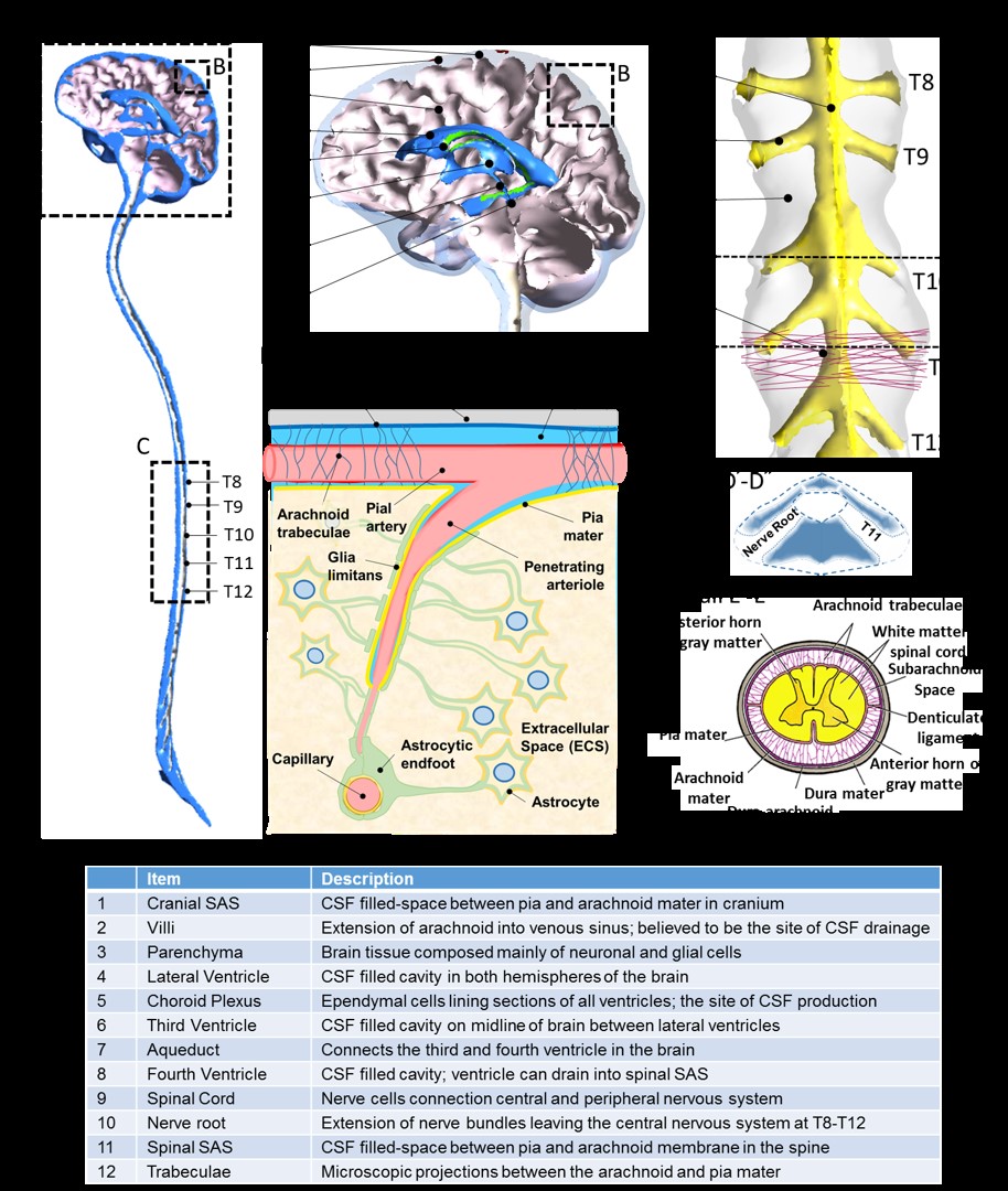
The brain and spinal cord are surrounded by CSF, a clear fluid with a density and viscosity close to water. It fills the ventricular system and the cranial and spinal SAS. CSF inside the CNS does not remain stagnant but pulsates 1. CSF also flows from the cranial to the spinal SAS in systole, with flow reversal from the spinal SAS into the cranium in diastole. A respiratory influence on the CSF oscillations in the aqueduct has also been observed 2–7. The exact source location and type of force coupling between the expanding and contracting vasculature, brain movement and induced CSF dynamics are still uncertain. Because the human CNS seems to lack a classical lymphatic system, CSF clearance differs from peripheral extracellular fluid drainage. CSF is believed to be reabsorbed into the venous system through the arachnoid villi, protrusions of the arachnoid membrane into the superior sagittal sinus, located (Figure 1a) or alternatively though nerve paths into the extracranial lymphatic system. Animal experiments suggest that lymphatic drainage is significant in rodents8,9 and dogs9. Recent findings point toward the existence of a meningeal lymphatic network in mice10. However, the extent of lymphatic drainage in humans is still debated11,12. Alternative theories of CSF production and reabsorption question this traditional view of CSF production 13–16. Supported by dilution experiments in cats, Klarica et al. (2005) proposed the theory that CSF production and reabsorption occur throughout the parenchyma without a clear bulk component due to choroidal production or clearance through arachnoid villi.Interstitial fluid dynamics.
The traditional view also holds that a fraction of CSF is secreted from the brain tissue, which is in agreement with Klarica et al.’s theory 17–19. New data suggest that the brain actively regulates the size of the ECS in accordance with metabolic needs 20. In addition to the possibility of fluid generation inside the tissue, ICF reabsorption from the interstitium into the microvasculature has been proposed as a way to interpret brain water content changes21. Experimental evidence also suggests the notion of net flow of CSF into the interstitial brain tissue 22. Several studies show that CSF can exit the ventricles under high pressure 23–25. Transependymal flow into perivascular spaces may be significant in hydrocephalus 26. Furthermore, the role of osmolarity exercising Starling forces needs to be quantified to determine the amount and directionality of bulk water exchange across the blood-brain barrier 27. Water exchange from astrocytic endfeet (Figure 1b) into the ECS sparked a growing interest in transmembrane proteins known as aquaporin channels 28–31. Despite these developments, a high degree of uncertainty about the amount, direction, and physiochemical driving forces of interstitial fluid exchange remains.Cerebrospinal Fluid Flow of the Spine.
In normal humans, 0.5-2 mL of CSF are displaced into the cervical SAS during systole in each cardiac cycle and flow back into the cranial SAS during diastole. Four-dimensional (4D) magnetic resonance measurements show a sharp CSF pulse shooting from the prepontine area into the cervical SAS 32. Additionally, flow velocities are higher in the anterior cervical SAS, with concentrated jets propagating along the cervical region. Craniocaudal flow into the spinal SAS requires concomitant expansions of the fluid-filled spaces. The systolic CSF inflow into the spinal SAS is believed to be accommodated by the deformation of the dura membrane, which in turn is enabled by displacement of venous blood or the compression of fatty epidural tissue especially in the lumbar region 33,34. PC MRI velocity measurements depict a gradual decline in the CSF stroke volume when measured at descending locations from the cervical to the lumbar spine. In addition to volumetric flow rate peak amplitude attenuation, a gradual increment in the phase lag of the velocity maximum was observed 35. Measured peak amplitude attenuation and phase lags were used to infer the volumetric strains responsible for spinal compliance 36. More precise measurements of the velocity waves and their timing are expected to localize and quantify the spatial extent of spinal dura deformations. The spinal CSF flow experiences intricate geometry-induced microflow patterns due to microanatomical aspects that can be found in the SAS (Figure 1c--e). Microanatomical features causing complex flows include ligaments, nerve roots, trabeculae, meningeal layers, and spinal white matter and gray matter, which have been carefully characterized by Reina et al. (2002a,b, 2004, 2015). Several groups are beginning to clarify the role of nerve roots on the geometry-induced CSF flow patterns in the spine37 36,38,39.Discussion
Research into CSF dynamics is an active field of research. Knowledge about CSF-ISF pathways is significant for a basic understanding of brain clearance functions that have relevance in brain pathologies (AD, Parkinsons). Moreover quantification of CSF dynamics is necessary for determining the driving forces of drug transport in non-invasive drug delivery modalities. These include intra-ventricular, intra-thecal and intra-parenchmal delivery options. Since many active drug molecules cannot cross the blood brain barrier, the significance of invasive drug delivery methods is expected to gain in importance.Conclusion
The overview raised open research questions as follows:
· What are the physiological connections between CSF and ISF.
· How are the fundamental mechanism of water homeostasis between the vascular and extravascular compartments? How is water exchanged in different brain compartments during awake and sleep stares as well as in normal and pathologies.
· What are physiological pathways and the fluid flows that are carried along lymph-like conduits along the venous sinuses that allow fluid to leave the cranial subarachnoid space without passing through the subarachnoid villi
· How are perivascular spaces connected to the lymph-like conduits
Acknowledgements
No acknowledgement found.References
1. Enzmann DR, Pelc NJ. Cerebrospinal fluid flow measured by phase-contrast cine MR. AJNR Am J Neuroradiol 1993;14:1301–1307; discussion 1309-1310.
2 Bhadelia RA, Madan N, Zhao Y, Wagshul ME, Heilman C, Butler JP, et al. Physiology-Based MR Imaging Assessment of CSF Flow at the Foramen Magnum with a Valsalva Maneuver. Am J Neuroradiol 2013;34:1857–1862. https://doi.org/10.3174/ajnr.A3509.
3 Dreha-Kulaczewski SF, Joseph AA, Merboldt K-D, Ludwig H-C, Gaertner J, Frahm J. Inspiration is the major regulator of human CSF flow. J Neurosci Off J Soc Neurosci 2015;35:2485–91. https://doi.org/10.1523/JNEUROSCI.3246-14.2015.
4 Kao Y-H, Guo W-Y, Liou AJ-K, Hsiao Y-H, Chou C-C. The respiratory modulation of intracranial cerebrospinal fluid pulsation observed on dynamic echo planar images. Magn Reson Imaging 2008;26:198–205. https://doi.org/10.1016/j.mri.2007.07.001.
5 Schroth G, Klose U. Cerebrospinal fluid flow. I. Physiology of cardiac-related pulsation. Neuroradiology 1992;35:1–9.
6 Williams B. Simultaneous cerebral and spinal fluid pressure recordings. Acta Neurochir (Wien) 1981;59:123–42. https://doi.org/10.1007/BF01411198.
7 Yamada S, Miyazaki M, Yamashita Y, Ouyang C, Yui M, Nakahashi M, et al. Influence of respiration on cerebrospinal fluid movement using magnetic resonance spin labeling. Fluids Barriers CNS 2013;10:36. https://doi.org/10.1186/2045-8118-10-36.
8 Leeds SE, Kong AK, Wise BL. Alternative pathways for drainage of cerebrospinal fluid in the canine brain. Lymphology 1989;22:144–6.
9 Zhao K, Sun H, Shan Y, Mao BY, Zhang H. Cerebrospinal fluid absorption disorder of arachnoid villi in a canine model of hydrocephalus. Neurol India 2010;58:371. https://doi.org/10.4103/0028-3886.65601.
10 Louveau A, Smirnov I, Keyes TJ, Eccles JD, Rouhani SJ, Peske JD, et al. Structural and functional features of central nervous system lymphatic vessels. Nature 2015;523:337–41. https://doi.org/10.1038/nature14432.
11 Wagshul ME, Johnston M. The brain and the lymphatic system. Immunol Lymphat Syst 2013:143–64. https://doi.org/10.1007/978-1-4614-3235-7_8.
12 Johnston M, Zakharov A, Papaiconomou C, Salmasi G, Armstrong D. Evidence of connections between cerebrospinal fluid and nasal lymphatic vessels in humans, non-human primates and other mammalian species. Cerebrospinal Fluid Res 2004;1:2. https://doi.org/10.1186/1743-8454-1-2.
13 Bulat M, Klarica M. Recent insights into a new hydrodynamics of the cerebrospinal fluid. Brain Res Rev n.d.;65:99–112. https://doi.org/10.1016/j.brainresrev.2010.08.002.
14 Klarica M, Miše B, Vladić A, Radoš M, Orešković D. “Compensated hyperosmolarity” of cerebrospinal fluid and the development of hydrocephalus. Neuroscience 2013;248:278–289. https://doi.org/10.1016/j.neuroscience.2013.06.022.
15 Bulat M, Lupret V, Orešković D, Klarica M. Transventricular and transpial absorption of cerebrospinal fluid into cerebral microvessels. Neurol Croat Suppl 2007;56:32–3.
16 Orešković D, Klarica M. A new look at cerebrospinal fluid movement. Fluids Barriers CNS 2014;11:16. https://doi.org/10.1186/2045-8118-11-16.
17 Milhorat TH, Hammock MK, Fenstermacher JD, Levin VA. Cerebrospinal fluid production by the choroid plexus and brain. Science 1971;173:330–332.
18 Milhorat TH. Choroid plexus and cerebrospinal fluid production. Science 1969;166:1514–1516.
19 Cserr HF. Physiology of the choroid plexus. Physiol Rev 1971;51:273–311. https://doi.org/10.1152/physrev.1971.51.2.273.
20 Iadecola C, Nedergaard M. Glial regulation of the cerebral microvasculature. Nat Neurosci 2007;10:1369–76. https://doi.org/10.1038/nn2003.
21 Penn RD, Bacus JW. The brain as a sponge: a computed tomographic look at Hakim’s hypothesis. Neurosurgery 1984;14:670–5.
22 Penn RD, Linninger A. The Physics of Hydrocephalus. Pediatr Neurosurg 2009;45:161–174. https://doi.org/10.1159/000218198.
23 Lebret K, Kritzberg ES, Rengefors K. Population Genetic Structure of a Microalgal Species under Expansion. PLOS ONE 2013;8:e82510. https://doi.org/10.1371/journal.pone.0082510.
24 Hodel J, Lebret A, Petit E, Leclerc X, Zins M, Vignaud A, et al. Imaging of the entire cerebrospinal fluid volume with a multistation 3D SPACE MR sequence: feasibility study in patients with hydrocephalus. Eur Radiol 2013;23:1450–8. https://doi.org/10.1007/s00330-012-2732-7.
25 Sæhle T, Eide PK. Intracranial pressure monitoring in pediatric and adult patients with hydrocephalus and tentative shunt failure: a single-center experience over 10 years in 146 patients. J Neurosurg 2015;122:1076–86. https://doi.org/10.3171/2014.12.JNS141029.
26 Hopkins LN, Bakay L, Kinkel WR, Grand W. Demonstration of transventricular CSF absorption by computerized tomography. Acta Neurochir (Wien) 1977;39:151–7. https://doi.org/10.1007/BF01406724.
27 Buishas J, Gould IG, Linninger AA. A computational model of cerebrospinal fluid production and reabsorption driven by Starling forces. Croat Med J 2014;55:481–97. https://doi.org/10.3325/cmj.2014.55.481.
28 Haj-Yasein NN, Jensen V, Vindedal GF, Gundersen GA, Klungland A, Ottersen OP, et al. Evidence that compromised K+ spatial buffering contributes to the epileptogenic effect of mutations in the human Kir4.1 gene (KCNJ10). Glia 2011;59:1635–42. https://doi.org/10.1002/glia.21205.
29 MacAulay N, Zeuthen T. Water transport between CNS compartments: contributions of aquaporins and cotransporters. Neuroscience 2010;168:941–56. https://doi.org/10.1016/j.neuroscience.2009.09.016.
30 Nedergaard M, Ransom B, Goldman SA. New roles for astrocytes: redefining the functional architecture of the brain. Trends Neurosci 2003;26:523–30. https://doi.org/10.1016/j.tins.2003.08.008.
31 Papadopoulos MC, Verkman AS. Aquaporin water channels in the nervous system. Nat Rev Neurosci 2013;14:265–77. https://doi.org/10.1038/nrn3468.
32 Bunck AC, Kröger J-R, Jüttner A, Brentrup A, Fiedler B, Schaarschmidt F, et al. Magnetic resonance 4D flow characteristics of cerebrospinal fluid at the craniocervical junction and the cervical spinal canal. Eur Radiol 2011;21:1788–1796. https://doi.org/10.1007/s00330-011-2105-7.
33 Shapiro K, Marmarou A, Shulman K. Characterization of clinical CSF dynamics and neural axis compliance using the pressure-volume index: I. The normal pressure-volume index. Ann Neurol 1980;7:508–14. https://doi.org/10.1002/ana.410070603.
34 Fig. (2). (From Marmarou et al. 1975). The volume-pressure response... ResearchGate. n.d. URL: https://www.researchgate.net/figure/Fig-2-From-Marmarou-et-al-1975-The-volume-pressure-response-test-for-invasive_fig2_233491320 (Accessed 30 April 2019).
35 Wagshul M, J Chen J, Egnor M, J McCormack E, E Roche P. Wagshul ME, Chen JJ, Egnor MR, McCormack EJ, Roche PEAmplitude and phase of cerebrospinal fluid pulsations: experimental studies and review of the literature. J Neurosurg 104:810-819. vol. 104. 2006.
36 Tangen KM, Hsu Y, Zhu DC, Linninger AA. CNS wide simulation of flow resistance and drug transport due to spinal microanatomy. J Biomech 2015;48:2144–2154. https://doi.org/10.1016/j.jbiomech.2015.02.018.
37 Pahlavian SH, Yiallourou T, Tubbs RS, Bunck AC, Loth F, Goodin M, et al. The Impact of Spinal Cord Nerve Roots and Denticulate Ligaments on Cerebrospinal Fluid Dynamics in the Cervical Spine. PLOS ONE 2014;9:e91888. https://doi.org/10.1371/journal.pone.0091888.
38 Stockman A, Plummer DJ, Montag ED. Spectrally opponent inputs to the human luminance pathway: slow +M and −L cone inputs revealed by intense long-wavelength adaptation. J Physiol 2005;566:61–76. https://doi.org/10.1113/jphysiol.2005.084046.
39 Hettiarachchi HDM, Hsu Y, Harris TJ, Linninger AA. The Effect of Pulsatile Flow on Intrathecal Drug Delivery in the Spinal Canal. Ann Biomed Eng 2011;39:2592. https://doi.org/10.1007/s10439-011-0346-x.
Figures