Advanced Brain MRI: Functions & Circuits
Joanes Grandjean1
1Singapore Bioimaging Consortium, Agency for Science, Technology and Research, Singapore, Singapore
Synopsis
Functional Magnetic Resonance Imaging in rodents open new avenues to understand the mechanisms underlying distributed neuronal networks and their dynamics in the healthy and diseased brain. Here, I will review the major observations and current limitations associated with the method. I will present the results from the first multi-center mouse resting-state fMRI comparison and will highlights the needs for the adoption of standards in the field.
A
comprehensive understanding of the architecture and function of the healthy and
diseased brain, often referred to as the “connectome”, is arguably one of the
biggest challenges in neuroscience.
Whole-brain analysis of structure and function using neuroimaging tools
provides valuable insight into information processing at the macroscopic level.
Functional imaging has been extensively used to map the healthy and diseased human
brain, to localize brain activity evoked by specific cognitive tasks or
estimate large-scale brain networks during rest. Advances in high field magnets
and radio-frequency coils now enable researchers to extend these studies to
animal models, where brain circuits can be further dissected using precise circuit
manipulation tools such as optogenetics. In this talk, I will detail the
development and considerations for functional connectivity analysis in mice and
its application in the field of affective disorder. Finally, I will expend on
the application of optogenetic together with functional magnetic resonance
imaging (ofMRI) to manipulate and visualize circuits in the living mouse. The
relevance of ofMRI is accentuated as it remains to-date the only available
method to visualize functional activity evoked with optogentics across the whole-brain
non-invasively. These studies offer a strong translational perspective to
investigate the molecular mechanisms behind MRI-based fingerprints of human
brain disorders, or to partake in the drug development process.
Acknowledgements
No acknowledgement found.References
No reference found.Figures
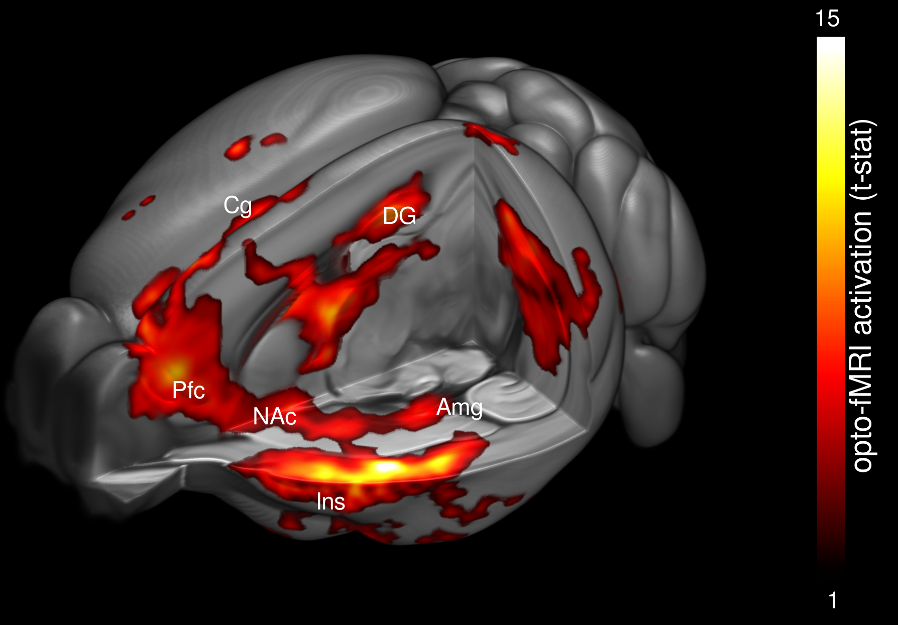
Optogenetic photostimulation of the mouse entorhinal cortex visualized with functional Magnetic Resonance Imaging