4691
Simultaneous Acquisition of Orthogonal Plane Cine Imaging and Isotropic 4D-MRI Using Super-Resolution1Radiation Oncology, Medical College of Wisconsin, Milwaukee, WI, United States
Synopsis
The super-resolution-based, isotropic 4D-MRI, and orthogonal cine imaging pulse sequence (SR-4D-SOPI) was developed and evaluated in this work. Cine imaging was acquired at more than 3 frames per second in sagittal and coronal planes simultaneously while also acquiring two orthogonal 4D-MRI volumes. The volumes were combined using super-resolution methods to create an isotropic 4D-MRI to be used for dose reconstruction following each fraction of abdominal or thoracic MR-guided radiation therapy.
Introduction
Real-time intrafraction motion monitoring of targets and organs at risk (OAR) is essential for abdominal and thoracic MR-guided radiation therapy (MR-gRT). Ideally, real-time volumetric information would be available for monitoring and reconstruction of the daily dose deposited to tumor and OAR in the presence of complex motions. The simultaneous orthogonal plane imaging (SOPI) method was developed to improve the accuracy of motion estimation during MR-gRT through time-locked sampling of orthogonal cine imaging planes[1]. An extension of SOPI, which simultaneously supports real-time prospective cine imaging and retrospective respiratory-correlated 4D-MRI generation for dose reconstruction was recently introduced[2]. We demonstrate here a further extension of SOPI that permits generation of an isotropic respiratory-correlated 4D-MRI volume using super-resolution (SR) reconstruction of two orthogonal, low-resolution (LR) 4D-MRI volumes.Methods
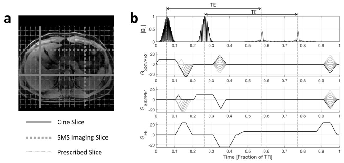
The super-resolution 4D simultaneous orthogonal plane imaging (SR-4D-SOPI) pulse sequence is shown in Figure 1. In each orthogonal axis, one slice remains in a constant position while the position of another slice along each axis is modulated from frame to frame. Signal from four slices is simultaneously acquired within each TR. The sagittal/coronal slice groups were separated via a simultaneous image refocusing (SIR) readout while the parallel slices were separated via the 2D-SENSE-GRAPPA parallel imaging algorithm[3].
An image-driven respiratory surrogate was derived from the cine images along both axes. The surrogate was used to retrospectively sort the concurrently acquired slices six respiratory phases. For each phase of the LR sagittal and coronal 4D-MRIs, a super-resolution reconstruction combining the orthogonal volumes was performed. The model matrix, A, maps the reconstructed isotropic data vector to the space of each orthogonal volume. It includes a geometric transformation to match orientations and origins of the acquired LR orthogonal volumes (G), blurring to model a Gaussian point spread function (B), and a down-sampling and cropping operator (D).
$$\mathbf{A_k} = \mathbf{D_k}\mathbf{B_k}\mathbf{G_k}$$
The SR reconstruction problem is posed as follows:
$$x = \underset{x}{\operatorname{argmin}} \sum_{k=1}^{N}\lVert \mathbf{y_k} - \mathbf{A_k}\mathbf{x} \rVert _2^2 $$
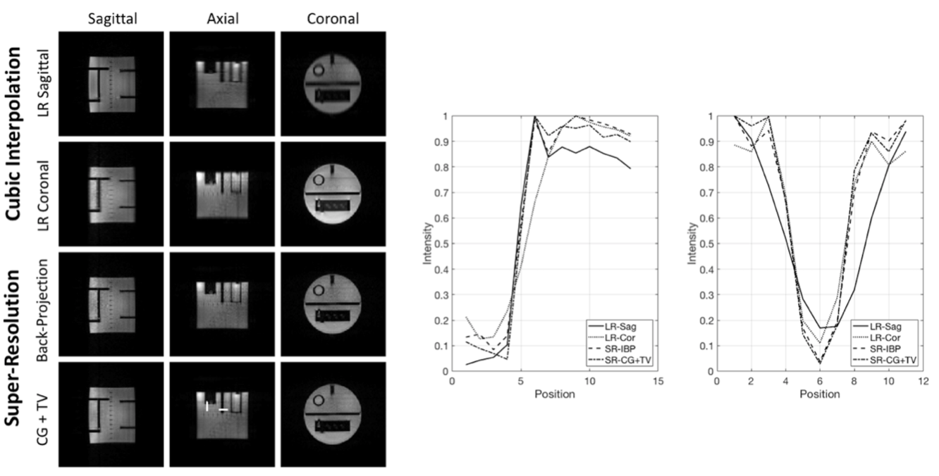
The solution to this problem was addressed with an iterative back-projection algorithm. A 3D total-variation-regularized solution was also found via conjugate gradient methods (CG+TV)[4,5].
The SR-4D-SOPI method with RF and gradient spoiling was implemented on a 3T Siemens Verio evaluated in phantom (for structural analysis) and in the abdomen of two consenting, free-breathing, liver cancer patients. The frame rate of the cine images for the in vivo scans was 3.23 frames per second. The mean structural similarity between the SR and the LR volumes were calculated relative to a separately acquired 3D high resolution (HR) scan.
Results
From the phantom experiment, it can be seen that the SR methods were able to produce isotropic volumes with high-resolution information in all three dimensions (See Figure 2). The line intensity profiles show sharp edges in the SR images matching the in-plane resolution in the LR volumes. The mean SSIM of the SR, LR sagittal, and LR coronal images relative to the HR images in the axial plane were 0.8016, 0.7017, 0.7249, respectively. An ANOVA analysis showed that the mean SSIM values between the SR, LR sagittal, and LR coronal volumes are significantly different (p<0.0001).
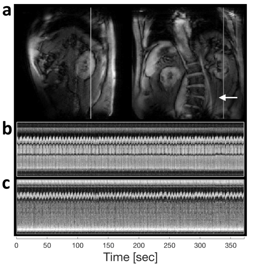
A single frame of orthogonal cine images is shown in Figure 3, along with a time series of intensity projections. Respiratory motion is adequately sampled within each cine imaging plane, facilitating the retrospective construction of LR orthogonal 4D-MRIs. The white arrow denotes residual aliasing due to imperfect parallel imaging separation of the SMS slices.
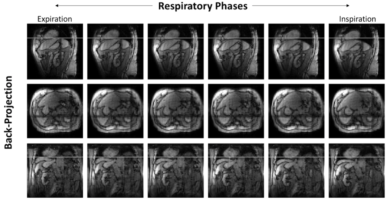
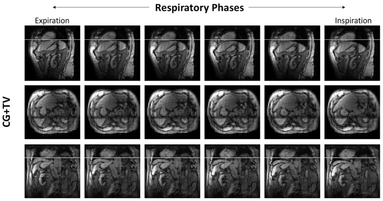
Figures 4 and 5 display the full isotropic SR reconstructions obtained with the back-projection and CG+TV algorithms, respectively. The white line was provided to visualize the degree of displacement of the dome of the liver as a function of respiratory phase. The CG+TV methods subtly reduces noise in the SR reconstructions compared with the back-projection reconstruction.
Discussion
A novel approach was presented that permits the simultaneous acquisition of cine and 4D-MRI imaging in two orthogonal planes. The cine imaging data serves two purposes. First, it can be used for prospective, real-time, intrafraction motion monitoring during MR-gRT. Second, it is used to retrospectively sort the concurrently acquired LR 4D-MRI slices into appropriate respiratory phase bins. The simultaneous acquisition of multi-respiratory phase 3D volumes enables retrospective, super-resolution reconstructions of isotropic 4D-MRI epochs.Conclusion
The SR-4D-SOPI method was demonstrated to provide cine imaging in two orthogonal planes while simultaneously providing low-resolution 4D-MRI in the same two orthogonal planes. A super-resolution reconstruction was able to successfully combine the two LR 4D-MRI datasets into one isotropic resolution 4D-MRI. Future work will explore the utility of SR-4D-SOPI for intrafraction motion management and truth-in-delivery analyses in MR-gRT.Acknowledgements
No acknowledgement found.References
[1] Mickevicius NJ, Paulson ES. Simultaneous orthogonal plane imaging. Magn Reson Med 2017;78:1700–10. doi:10.1002/mrm.26555.
[2] Mickevicius NJ, Chen X, Boyd Z, Lee HJ, Ibbott GS, Paulson ES. Simultaneous motion monitoring and truth-in-delivery analysis imaging framework for MR-guided radiotherapy. Phys Med Biol 2018. doi:10.1088/1361-6560/aaec91.
[3] Koopmans PJ. Two-dimensional-NGC-SENSE-GRAPPA for fast, ghosting-robust reconstruction of in-plane and slice-accelerated blipped-CAIPI echo planar imaging. Magn Reson Med 2016. doi:10.1002/mrm.26179.
[4] Plenge E, Poot DHJ, Bernsen M, Kotek G, Houston G, Wielopolski P, et al. Super-resolution methods in MRI: Can they improve the trade-off between resolution, signal-to-noise ratio, and acquisition time? Magn Reson Med 2012;68:1983–93. doi:10.1002/mrm.24187.
[5] Van Reeth E, Tan CH, Tham IW, Poh CL. Isotropic reconstruction of a 4-D MRI thoracic sequence using super-resolution. Magn Reson Med 2015;73:784–93. doi:10.1002/mrm.25157.
Figures