4349
A navigator-guided novel motion correction method in simultaneous PET/MR abdominal imaging1Department of Nuclear Medicine, Zhongshan Hospital, Shanghai, China, 2United Imaging Healthcare, Shanghai, China, 3UIH America, Houston, TX, United States
Synopsis
Simultaneous PET/MR abdominal imaging suffers from respiratory artifacts which causes motion blur of the PET images, further affecting clinical diagnosis. To eliminate motion blur, the work presents a novel PET image motion correction method by optimizing attenuation maps with navigators. The method significantly improves the image quality as well as the work flow and demonstrates its clinical value in detecting lesion in the liver.
Purpose
The work presents a novel PET image motion correction method by optimizing attenuation maps with navigators (NavMC) in simultaneous PET/MR abdominal imaging. The technique demonstrates its capability in eliminating motion blur and its clinical application value in detecting lesion in the liver region which are susceptible to motion artifacts.Introduction
Simultaneous PET/MR abdominal imaging suffers from respiratory artifacts which causes motion blur of the PET images, further affecting clinical diagnosis.1 To eliminate motion blur, conventional motion correction methods involve using respiratory motion signals recorded by external devices (e.g. belts).2 However, these methods are complex in patient positioning and lack of accuracy in measuring organ movement. By taking the advantages of the state-of-art PET/MR system, in this work, we propose to use simultaneously acquired MR navigators to directly track the motion of liver. Additionally, we present a novel PET image motion correction method by optimizing attenuation maps to correct the blurring artifacts.Methods
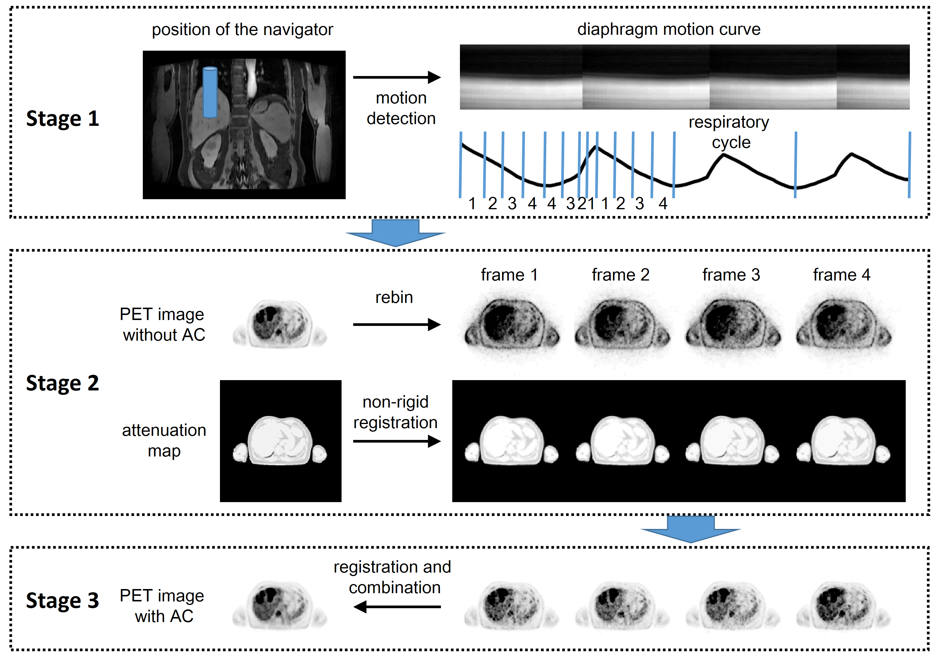
The experiments were performed on a simultaneous whole-body PET/MR scanner (Unites Imaging, Shanghai, China) in Zhongshan Hospital, Shanghai, China. The PET image acquisition was performed in clinical settings in conjunction with routine MR scans for diagnosis purposes. The procedures of PET image reconstruction with the NavMC method are summarized in Figure 1:
- (a) Position a real-time 2D excitation pencil beam navigator at the right hemi-diaphragm to monitor respiratory motion and use the motion signal to trigger PET/MR image acquisition; (b) Extract respiratory motion waveform by tracking the location of liver using an edge detection algorithm and separate the waveform to 4 time phases according to motion amplitude in a respiratory cycle.
- (a) Rebin the acquired PET image data to 4 frames based on the time windows and reconstruct them individually; (b) Transform an attenuation map acquired with no MC to each frame of PET images using a non-rigid registration algorithm.
- Reconstruct each frame of PET image using transformed attenuation maps and summed to generate a high SNR PET image.
The performance of the proposed NavMC method was evaluated compared to: (1) no motion correction (NoMC); (2) motion correction using respiratory motion signals acquired by a chest belt placed around abdomen (ResMC). For clinical evaluation, image acquisitions were performed on five patients who underwent routine clinical 18F-FDG PET scans with different motion correction methods.
Results
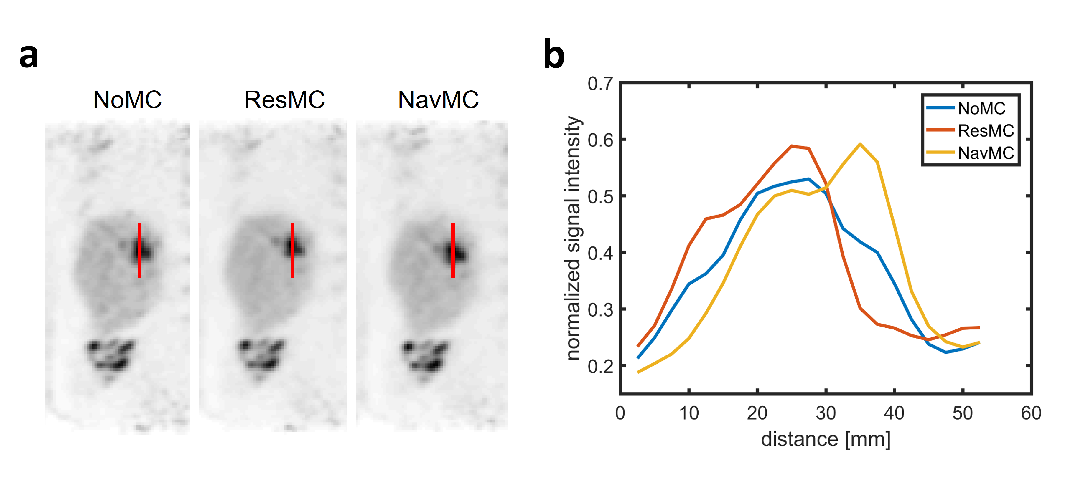
The boxplot in Figure 2 compares signal distribution across the lesion with NoMC, ResMC, and NavMC. As can been seen, NavMC yields a reduction in image blur in lesion, while delivering higher peak signal than the NoMC method. ResMC shows a similar pattern of signal distribution as NavMC.
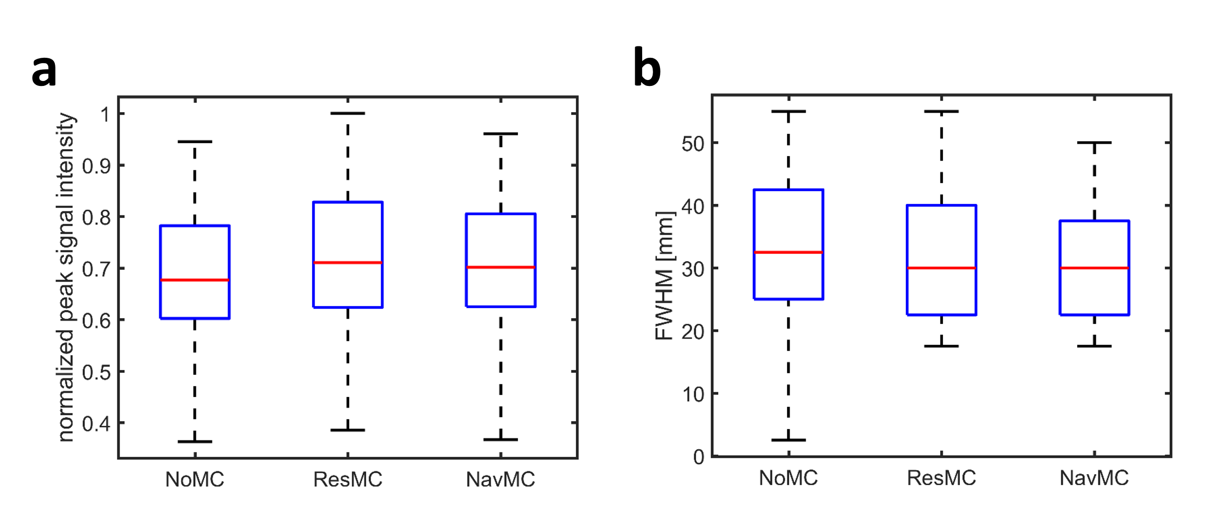
Figure 3 shows the experimental results on five example subjects. The boxplot (a) and (b) show the Full Width at Half Maximum (FWHM) value of lesions area in the liver and the corresponding peak signal in multiple locations, respectively. NoMC, ResMC, and NavMC provide an average signal peak of 0.68, 0.71 and 0.7 and FWHM by 33mm, 30mm, and 30mm. The boxplot (b) shows both NavMC and ResMC yield a reduction in standard deviation than NoMC.
Discussion
In simultaneous PET/MR imaging, the NavMC method significantly improves the image quality as well as the work flow since it requires no external vital signal monitoring devices. It minimizes the motion effect in PET/MR without loss of signal to noise ratio. The accuracy and speed of optimization can be further improved by using advanced machine learning based non-rigid registration methods.Conclusion
The NavMC method yields better results as the NoMC method and comparable results as the ResMC method. Meanwhile, this method demonstrates its clinical value in detecting lesion in the liver.Acknowledgements
No acknowledgement found.References
1. Liu C, Pierce II LA, Alessio AM , et al. The impact of respiratory motion on tumor quantification and delineation in static PET/CT imaging. Phys Med Biol. 2009;54(24):7345.
2. Klein GJ, Reutter BW, Ho MH, et al. Real-time system for respiratory-cardiac gating in positron tomography. IEEE Trans Nucl Sci. 1998;45(4):2139–2143.
Figures