4272
In vivo Fast Field Cycling Relaxometry reports on the extra- and intracellular localization of iron oxide particles in tumour mice models.1University of Torino, Torino, Italy
Synopsis
A new sensitive, non invasive, imaging methods capable of a quantitative detection of Tumour Associated Macrophages (TAM) is herein proposed. The method is based the use Fast Field Cycling (FFC) relaxometers to assess the localization of ferumoxytol in TAM in melanoma tumours. The observed 1/T1 NMRD profiles appear highly dependent on the intra or extra cellular localization of the NPs thus allowing an unambiguous TAM quantification.
Introduction
The complex relationships between immune system and tumour is under intense scrutiny as they are considered an important hallmark of cancer1. Tumour associated macrophages (TAMs) are forced by the cancer cells to adopt an anti-inflammatory phenotype and secrete factors to promote angiogenesis and tumor invasion. For these reasons, sensitive, non invasive, imaging methods that are capable of a quantitative detection of TAM are needed. To this purpose, the use of Ultra Small Iron Oxides nanoparticles (NPs) has been already proposed because they are taken up by TAM generating a detectable contrast in T2 weighted images. However, the main drawback of this approach relies on the fact that the observed contrast is not dependent on the extra- or intracellular localization of the NPs. Thereafter, there is the need to develop new methods able to discriminate the intra and extra-cellular localization of NPs only for TAM detection and, in general, for a number of “cell tracking” applications. To this purpose we have tested the use Fast Field Cycling (FFC) relaxometers to assess the localization of ferumoxytol (a clinical approved USPIO NPs) in TAM in melanoma tumours. The observed NMRD profiles appear highly dependent on the intra or extracellular localization of the NPs thus allowing an unambiguous TAM quantification.Methods
NMRD profiles were acquired on a FFC relaxometer able to switch the magnetic field over a large range of field stranghts (0.01-20MHz). The application of a FFC overcomes the problem of the low sensitivity at low magnetic fields and allows rapid acquisition of the magnetization recovery at each magnetic field strength. In order to host a mouse, the commercially available relaxometer (Stelar, Mede, Italy) has been modified with the implementation of a 40 mm 0.5T Field Cycling magnet and a dedicated 11mm solenoid detection coil placed around the anatomical region of interest2. The tumour xenografts were prepared by injecting three tumour cell lines (B16 melanoma, 4T1 and 168FARN breast carcinoma) in the hindlimb muscle.Results
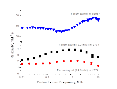
First in vitro studies have been carried out on a murine monocyte-derived macrophage cell line (J774) to evaluate the relaxivity changes due to the intracellular localization of ferumoxytol. The NMRD profile of ferumoxytol labelled J774 cells (Figure 1) differs markedly from the one obtained from ferumoxytol suspended in aqueous solution. In fact, the relaxivity peak at ca. 8-10 MHz observed in water is shifted to lower magnetic field strengths (at 0.5-1 MHz) when the NPs were entrapped in macrophages). Furthermore, the relaxivity values of the entrapped NPs were significantly lower than those observed in water. Likely, this is the consequence of the occurrence of a substantial relaxivity “quenching” when the magnetic particles are compartmentalized in intracellular vesicles such as endosomes or lysosomes. On the basis of these observations, the NMRD profiles of three different tumour cell lines were acquired in vivo. The selected types of tumours (168FARN, 4T1 and B16) are characterized by different amount of necrotic zones and macrophages infiltrating the tumor stroma. Ferumoxytol was injected at a dose of 0.5 mmol/kg of Fe. The profile obtained 3h and 24h after the injection were significantly different. (Figure 2) The profile observed at 24h displays a bell-shaped profile with a maximum around 0.4-0.5 MHz similar to one found for ferumoxytol labelled J774 macrophages. This finding clearly indicated the intracellular localization of ferumoxytol as confirmed by histological analysis by the Pearls assay.Conclusion
From these results, one can conclude that the characteristics of the NMRD profile immediately reports on the intra- or extra-cellular localization of the investigated contrast agent (ferumoxytol). This information could be hardly achievable from the measurements at single magnetic field and open new horizons for the field of cell tracking applications. Despite the herein used prototype FFC-NMR instrumentation is not endowed with spatial resolution, FFC has recently been applied to MRI, largely thanks to the work of the Lurie group at Aberdeen University where two prototype human whole-body sized FFC-MRI scanners have been built3.Acknowledgements
This project has received funding from the European Union’s Horizon 2020 research and innovation programme under grant agreement No 668119References
[1] Morita Y, Zhang R, Leslie M, Adhikari S, Hasan N, Chervoneva I, Rui H, Tanaka T. Oncol Lett. 2017;14:2111-2118.
[2] Ruggiero MR, Baroni S, Pezzana S, Ferrante G, Geninatti Crich S, Aime S. Angew Chem Int Ed Engl. 2018, 57:7468-7472.
[3] Pine KJ, Davies GR, Lurie DJ. Magn Reson Med., 2010, 63, 1698-702.
Figures