4251
Proton MRS and resting state fMRI study of hyperbaric oxygenation effects at 3 Tesla1Emanuel Institute of Biochemical Physics of RAS, Moscow, Russian Federation, 2Radiology, Clinical and Research Institute of Emergency Pediatric Surgery and Traumatology, Moscow, Russian Federation, 3Semenov Institute of Chemical Physics of the Russian Acedemy of Sciences, Moscow, Russian Federation, 4Clinical and Research Institute of Emergency Pediatric Surgery and Traumatology, Moscow, Russian Federation
Synopsis
Previously we demonstrated that one hyperbaric oxygenation (HBO) session causes the energy metabolism activation in order to compensate the expenses on NAD(H) synthesis. This study is aimed to reveal the effects of HBO on the 1H MRS brain metabolites and the correlated activity in DMN at 3 Tesla. The results demonstrate that even one low-pressure HBO session in healthy subjects stimulates NAA brain metabolism and increases the MPFC-PCC functional connectivity. Further research of HBO effects on neuronal pathology subjects is of interest.
Introduction
Hyperbaric oxygenation (HBO) has proved itself as an effective way of treatment in different cases, for example in hypoxia1. However, the exact in vivo biochemical mechanisms of HBO are to be revealed. Previously using 31P MRS we have found that one hyperbaric oxygenation (HBO) session causes the energy metabolism activation in order to compensate the expenses on NAD(H) synthesis2,3. Also we have demonstrated3 some promising preliminary results of 1H MRS metabolite concentration changes and resting state fMRI (rsfMRI) connectivity on a small group of subjects. Now we have enlarged the group of subjects and measured the absolute concentrations of 1H MRS metabolites in order to obtain more accurate and statistically significant results.Materials and methods
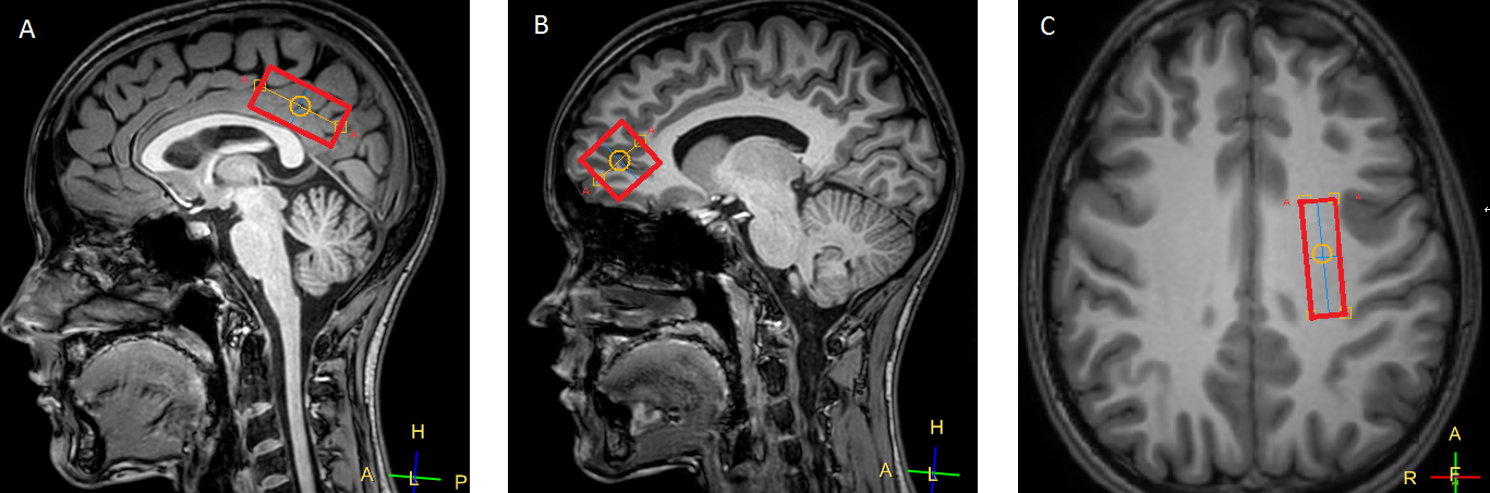
Philips Achieva dStream 3.0T, Philips Head 32-channel SENSE coil and hyperbaric chamber Sechrist 3200 were used. Twelve normal subjects (aged 18-30) participated in the study. At first, the survey images and 3D T1w images were obtained. After that, the resting state fMRI (GE-EPI, TR=3000 ms, TE=30 ms, EPI factor = 240, slice 4 mm, matrix = 160x160, 80 dynamic scans) and 3 single-voxel 1H MRS (PRESS, TE = 115 ms, TR = 2000 ms) were performed. Voxels were located in MPFC (25x25x25 mm), in PCC (40x20x20 mm) and in the left subcortical white matter (35x10x10), see fig.1. In order to maintain the equality of voxel location before and after HBO the 3D T1w images were positioned with extreme accuracy, rotation and slice location of voxels were saved before HBO and applied after HBO. Then subject proceeded to the HBO department. The HBO session lasted for 50 minutes (5 minutes for achieving constant 1.2 atmospheres mode, 40 minutes at the mode and 5 minutes for decompression). The time required for a patient to get to MRI scanner after HBO did not exceed 5 minutes. After HBO session the subject returned to the scanner, the where MRS and fMRI studies were repeated.Data processing
The compound of 1H MRS voxels was segmented on grey matter (GM), white matter (WM) and corticospinal fluid (CSF) fractions using 3D T1w images in SpectroComposition (the MATLAB tool for spectral quantification by Guillaume Gilbert). For each voxel the GM/WM/CSF fractions after HBO were normalized on the corresponding values before HBO in order to find relative changes. These values were compared with the value=1 using Mann-Whitney criterion (MW).
The 1H MRS data was processed in LCModel, where the metabolite concentrations relative to unsuppressed H2O were obtained in spectra before and after HBO. In order to avoid the mistakes caused by the different spectral preparation parameters (e.g. different shimming before and after HBO) the absolute concentrations of metabolites ([NAA], [Cr], [Cho], [mI], and [Glx]) were calculated according to4, using the voxel segmentation data. For every voxel location the metabolite concentrations before HBO were normalized on the corresponding concentrations after HBO, these values were compared with the value=1 (MW).
The rsfMRI data was processed in CONN (MATLAB). The seed-based correlation analysis defined the functional connectivity of the default mode network (DMN) nodes. The Fisher correlation coefficients (β) between different DMN nodes before HBO were compared with the corresponding values after HBO (MW).
Results
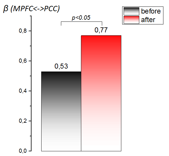
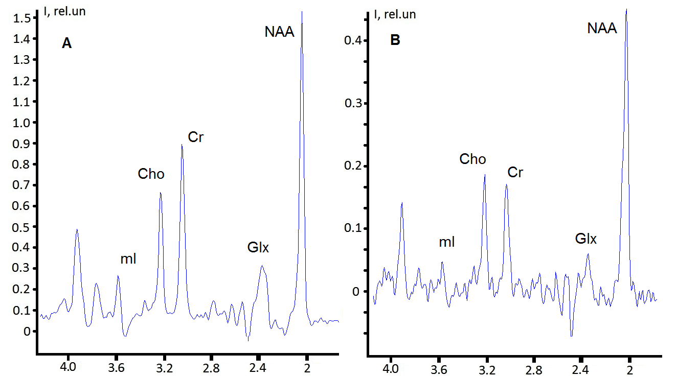
No changes in GM/WM/CSF fractions of all voxels were found after HBO. The β (MPFC<->PCC) increased after HBO, see fig 2. No changes found in β between other DMN nodes. Typical spectra obtained in this study are demonstrated on figure 3. Only [NAA] and only in MPFC was changed (p<0.05, decrease by 4%) after HBO, other concentrations remained unchanged.Discussion
No changes in voxel segmentation after HBO indirectly witnesses for the equality of voxel positioning, which means that all changes possible to be found are caused by metabolic and not partial-volume effects. The connectivity in DMN is linked with the core cognition processes, DMN connections are impaired in case of, for example, Alzheimer disease and post-traumatic stress syndrome5. The increase of β (MPFC<->PCC) after HBO is the direct improvement of DMN connectivity, that may help to overcome the negative effects of neurological disorders on brain functioning.
The functional connectivity activation after HBO has be connected with the increased energy expenses that we demonstrated in2,3. This also could be the reason for the decrease of NAA in MPFC, since NAA is reported to be involved in energy exchange6. The behavior of NAA and β (MPFC<->PCC) after HBO might be the manifestation of metabolism<->functioning connection. To sum up, the demonstrated results are the confirmation of HBO effectiveness for human brain stimulation even at low O2 pressures. Further research of HBO effects on human brain in case of neurological pathology is of interest.
Acknowledgements
No acknowledgement found.References
1. G.G. Rogatsky, S. Meilin, N. Zarchin, S.R. Thom, A. Mayevsky. Hyperbaric oxygenation affects rat brain function after carbon monoxide exposure. Undersea Hyperb Med. 2002 Spring;29(1):50-8
2. Manzhurtsev, A.V., Vasiukova, O.R., Sergeeva, V.V. et al. 31P MRS Study of Hyperbaric Oxygenation Effects on Phosphate Metabolism of Normal Human Brain. Appl Magn Reson (2018) 49: 679. https://doi.org/10.1007/s00723-018-1019-9
3. Andrei Manzhurtsev, Olga Vasiukova, Dmitry Kupriyanov, Victoria Sergeeva, Tolib Akhadov, Petr Menshchikov and Natalia Semenova. Multimodal study of hyperbaric oxygenation effects on normal human brain metabolism and functional connectivity at 3 Tesla. Proc.Int.Soc.Magn.Reson.Med (2018) #3273
4. Minati L, Aquino D, Bruzzone MG, Erbetta A. Quantitation of normal metabolite concentrations in six brain regions by in-vivoH-MR spectroscopy. J Med Phys. 2010;35(3):154-63.
5. Akiki TJ, Averill CL, Wrocklage KM, Scott JC, Averill LA, Schweinsburg B, Alexander-Bloch A, Martini B, Southwick SM, Krystal JH, Abdallah CG. Default mode network abnormalities in posttraumatic stress disorder: A novel network-restricted topology approach. Neuroimage. 2018 Aug 1;176:489-498
6. Manzhurtsev, A.V., Semenova, N.A., Ublinskii, M.V. et al. The effect of neurostimulation on the
intracellular concentrations of proton-containing metabolites and
macroergic phosphates in the brain cortex upon schizophrenia according
to the data from 1H and 31P magnetic resonance spectroscopy. Russ Chem Bull (2016) 65: 1630.
Figures