4056
Feasibility of free-breathing T1-weighted 3D radial VIBE for fetal MRI in various anomalies1Department of Radiology, International Peace Maternity and Child Health Hospital, School of Medicine, Shanghai Jiao Tong University, Shanghai, China, 2Diagnostic Imaging, SIEMENS Healthcare, Shanghai, China, 3R&D, Diagnostic Imaging, SIEMENS Healthcare, Erlangen, Germany
Synopsis
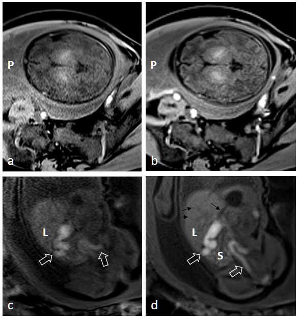
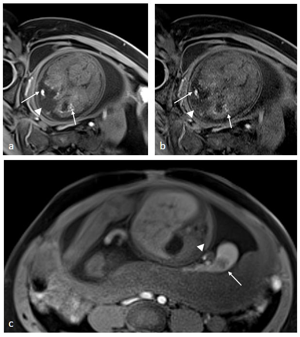
Traditional breath-hold, fat-suppressed T1-weighted sequences (T1W images) may be degraded by motion artifacts arising from both the mother and fetus. Images obtained with the radial VIBE sequence had a higher overall image quality score than Cartesian sampled 3D VIBE T1W images and less motion artifact in various fetal regions. Fetal lesions presented in radial VIBE images had good lesion conspicuity and edge sharpness.
Introduction
Traditional breath-hold, fat-suppressed T1-weighted sequences (T1W images) may be degraded by motion artifacts arising from both the mother and fetus.1 The T1-weighted 3D prototype radial VIBE sequence is reported to be a viable alternative to conventional Cartesian acquisition for pediatric MR in various settings, with better image quality.2-4 The purpose of this study was to evaluate the role of radial VIBE in multiple settings, including fetal head and neck, body and intra-uterus structures, compared with standard breath-hold T1W images, and its feasibility for demonstrating various fetal anomalies.Methods
A total of 248 pregnant women with a mean gestational age of 27.3 (range: 17 - 41.6) weeks who underwent fetal MRI were included. All the examinations were performed on a 1.5 T MR scanner (MAGNETOM Aera, Siemens Healthcare, Erlangen, Germany). The conventional Cartesian-sampled T1W sequence was performed under breath-holding conditions or in free breathing if the patient could not suspend respiration. The prototypical 3D stack-of-star radial VIBE sequence was subsequently used for all patients in free-breathing. The parameters of the conventional Cartesian-sampled 3D VIBE T1W sequence were: TR= 4.49 ms; TE= 2.19 ms; matrix size= 320 x 165; FOV, 380 x 261 mm2; slice number 30, and slice thickness, 3 mm. The parameters of the 3D radial VIBE sequence included: TR= 3.58 ms; TE= 1.69 ms; radial views= 800; matrix size= 256 x 256; FOV= 385 x 385 mm2; slice number 30, and slice thickness, 2.5 mm. Image quality and lesion conspicuity were evaluated by two radiologists blinded to the acquisition schemes on a five-point scale, with a higher score indicating a better trajectory method. Mixed-model analysis of variance and an interobserver variability assessment were performed.Results
Images obtained with the radial VIBE sequence had a higher overall image quality score than the T1W images (3.82 ± 0.95 vs. 2.88 ± 0.8, p < 0.001) and less motion artifact (p < 0.001). The radial VIBE images showed better tissue contrast (p < 0.001) and tissue edge clarity (p = 0.001) in the head and neck region. In the fetal body region, the radial VIBE sequence also showed a significantly better performance than that of conventional T1W imaging, including the overall image quality (3.8 ± 0.94 vs. 3.15 ± 0.87, p < 0.001), hepatic vessel clarity (p < 0.001), intestinal conspicuity (p < 0.001), and vessel clarity (p < 0.001). Good radial VIBE image quality was observed in the placenta and intra-uterine structures compared with the images acquired by conventional T1W images (4.17 ± 0.63 vs. 3.13 ± 0.72, p < 0.001). Of 49 lesions, 48 presented on radial VIBE images with good lesion conspicuity (4.54 ± 0.62) and edge sharpness (4.27 ± 0.76). The performance of radial VIBE for fetal lesion conspicuity by lesion location is listed in Table 1. A few streaking artifacts were found on radial VIBE images.Discussion
Our results demonstrate that clinical radial VIBE of the fetus is feasible during free breathing and that overall image quality is significantly better when compared with conventional breath-hold T1W images. Preliminary findings from Chandarana et al.5 showed superior image quality of the radial technique compared with standard Cartesian post-contrast imaging in free-breathing patients, and image quality that was at least comparable to that with breath-hold image acquisition. Our study demonstrated a similar result. Consistent with previous reports on pediatric MRI2-4, the results showed better image quality and lesion conspicuity in fetal head, neck, and body (p < 0.001). Intrauterine structures including the placenta and vessels could easily be affected by movement of adjacent maternal and fetal parts. By reducing multiple artifacts, radial VIBE also presented with significantly better placental vessel clarity and placental tissue contrast (p < 0.001). However, there remained a few lesions that scored less than 2 or even absent with radial VIBE (Table 1). This might be because of the intense fetal movement during the relatively long acquisition time of radial VIBE.Conclusion
For fetal imaging, the radial VIBE sequences had better image quality and lesion conspicuity than conventional breath-hold fat-suppressed T1-weighted sequences.Acknowledgements
No acknowledgement found.References
[1] Malamateniou C, Malik SJ, Counsell SJ, et al. Motion-compensation techniques in neonatal and fetal MR imaging. AJNR Am J Neuroradiol. 2013 Jun-Jul;34(6):1124-36.
[2] Park JE, Choi YH, Cheon JE,et al. Three-Dimensional Radial VIBE Sequence for Contrast-Enhanced Brain Imaging: An Alternative for Reducing Motion Artifacts in Restless Children. AJR Am J Roentgenol. 2018 Apr;210(4):876-882.
[3] Cho HH, Choi YH, Cheon JE, et al. Free-Breathing Radial 3D Fat-Suppressed T1-Weighted Gradient-Echo Sequence for Contrast-Enhanced Pediatric Spinal Imaging: Comparison With T1-Weighted Turbo Spin-Echo Sequence. AJR Am J Roentgenol. 2016 Jul;207(1):177-82.
[4] Chandarana H, Block KT, Winfeld MJ, et al. Free-breathing contrast-enhanced T1-weighted gradient-echo imaging with radial k-space sampling for paediatric abdominopelvic MRI. Eur Radiol. 2014 Feb;24(2):320-6.
[5] Chandarana H, Block TK, Rosenkrantz AB, et al. Free breathing radial 3D fat-suppressed T1-weighted gradient echo sequence: a viable alternative for contrast-enhanced liver imaging in patients unable to suspend respiration. Investig Radiol 46:648–653
Figures