3888
Dynamic Functional Connectivity Distinguishes Concussed Youth Based on Symptomology1University of Calgary, Calgary, AB, Canada
Synopsis
Concussion in youth is a growing health issue, and accurate diagnostic, progostic and assessment tools are lacking. In this study, we used a toolbox recently developed by our group (HOCo) to investigate dynamic functional connectivity in fMRI datasets obtained from groups of concussed and orthopedic injured (OI) control subjects. We estimated the proportion of the time that regions of the brain are significantly connected within specific networks. We found that concussed youth with somatic symptoms exhibited more stable connections of the motor and auditory networks, relative to OI subjects and concussed youth with cognitive symptoms.
Introduction
Concussion or mild traumatic brain injury (mTBI) is a growing health concern, and objective diagnosis and assessment remains lacking1, complicating clinical management. Previous MRI findings regarding structural and functional alterations in the brains of concussed individuals are highly variable. Examining the dynamic nature of brain functional connections is of increasing interest in the neuroimaging field; however, approaches to date are limited and cannot determine the significance of connections. Recently, we introduced an approach based on hierarchical observation modeling2, which allows the calculation of a t-statistic for connectivity between any windowed segment of two time-series, and, thus significance can be estimated. This method is now implemented in the Hierarchical Observation Connectivity (HOCo) toolbox, and was used in the present study to assess fMRI datasets of concussed and orthopedic-injured (OI) control subjects.Methods
In this multicenter study, concussed youth and OI controls were assessed using the Health and Behavior Inventory (HBI)3, and groups (N=13) were formed based on symptomology (somatic symptoms, cognitive symptoms, OI). All subjects underwent a 3-Tesla, 8-minute resting-state fMRI scan (TE/TE = 2000/30 ms; 3.6-mm isotropic voxels; matrix 64x64x36). Images were preprocessed using FSL4 following standard steps, and were then analyzed using HOCo. Six atlas-defined resting-state networks5 were selected for analysis: auditory (AUD), motor (MOT), left executive (LE), right executive (RE), default mode (DMN) and salience (SAL). The spatial mean time-series using (minimally modified) regions of interest (ROI) from these networks were extracted, and t-values as a function of time were calculated for all possible ROI pairs, using HOCo. The percentage of time that the t-value exceeded 2 was calculated as functional connectivity endurance (FCE), which was then compared between groups using Fisher’s exact test.Results
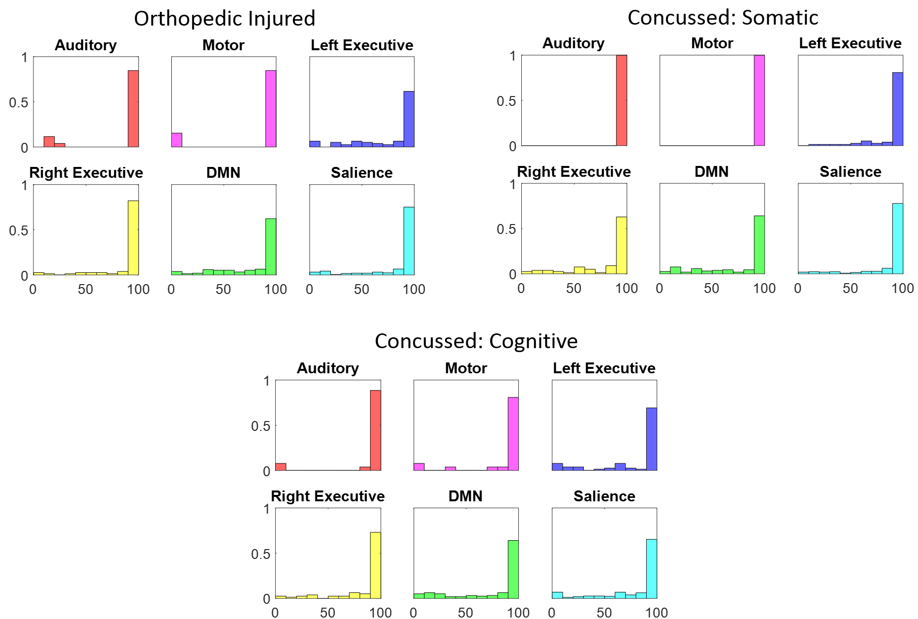
FCE histograms for all groups are shown in Figure 1 for within-network functional connections. As there were a large number of connections close to 100% for all networks, we use Fisher’s exact test to compare the proportion of connections close to 100% (last to bin to the right of all histograms) against all the others combined (remaining bins) within each network. After Bonferroni correction for multiple comparisons, in comparison to the somatic symptom group, significant differences (p< 0.05) were found for the auditory (OI vs. Somatic: p=0.0018; Cognitive vs. Somatic: p=0.0054) and motor networks (OI vs. Somatic: p=0.0018; Cognitive vs. Somatic: p=0.0018).Discussion
FCE, as calculated using HOCo, is able to distinguish functional network alterations of concussed individuals with differing symptomologies. Somatic symptoms are associated with more stable connections within motor and auditory netowrks, perhaps as a means to compensate for somatic symptoms. Longitudinal studies are thus necessary to ascertain whether FCE normalizes as symptoms resolve.Conclusion
Functional connectivity endurance (FCE) based on hierarchical observational modelling of dynamic connectivity holds promise as a technique to more accurately describe the impact of concussion on the youth brain. Further studies are planned to examine changes in FCE over time, and to determine if FCE has prognostic capabilities for functional outcomes.Acknowledgements
No acknowledgement found.References
1. Yeates KO, Beauchamp M, Craig W, et al. Advancing Concussion Assessment in Pediatrics (A-CAP): a prospective, concurrent cohort, longitudinal study of mild traumatic brain injury in children: protocol study. BMJ Open. 2017;7(7):e017012.
2. Sojoudi A, Goodyear BG. Statistical inference of dynamic resting-state functional connectivity using hierarchical observation modeling. Hum. Brain Mapp. 2016;37(12):4566-4580.
3. Ayr LK, Yeates KO, Taylor HG, et al. Dimensions of postconcussive symptoms in children with mild traumatic brain injuries. J. Int. Neuropsychol. Soc. 2009;15(1):19-30.
4. Jenkinson M, Beckmann CF, Behrens TEJ, et al. FSL. 2012;62(2):782-90.
5. Thomason ME, Dennis EL, Joshi AA, et al. Resting-state fMRI can reliably map neural networks in children. Neuroimage. 2011;55(1):165-75.
Figures