3634
Taxon anisotropic phantom delivering human scale parametrically controlled diffusion compartments to advance cross laboratory research and calibration.1University of Pittsburgh, Pittsburgh, PA, United States, 2Psychology Software Tools, Pittsburgh, PA, United States
Synopsis
Details a novel bi-component polymer Taxon (textile water filled tubes) anisotropic diffusion phantom providing 0.8 microndiameter tubes, packing density of 106 per mm2, water fraction of over 45% matched to human axon histology with parametric control of water compartment (intra/extra axon), size, density, angle dispersion that can be manufactured to supply hundreds of laboratories. Observed human tissue range FA (0.6-01.0), MD and RD in observed range. Providing ground truth measurement to advance and calibrate anisotropic diffusion measurement. Micro-CT and diffusion MRI indicate high water content in agreement with MRI and non MRI measurements.
INTRODUCTION
Since the invention of diffusion imaging over 30 years ago there has been an unmet need for “ground truth” measurement at the scale of human axonal tissue. By advancing state of the art of textile manufacturing, we can deliver parametric control of diffusion axonal chambers. We can produce reference phantoms using Taxons (textile water filled tubes the size of axons) meeting the demanding specification of providing .8 micrometer average diameter tubes with a packing density of 106 per mm2 with a water fraction of over 45% and provide large quantities of fibers (tens of kilometers long). These dimensions are based on histology measurements from modal human, chimp and monkey corpus callosum axon samples [1]. There have been a series of past anisotropic phantoms [2-15] but none have broken through the 5 micron internal diameter limit. This project introduces novel sub-micron fabrication methods that can fully cover the range of human tissue with parametric control of micro compartment water chambers. This project introduces a new technology into MRI phantoms of bi-component polymer textile production. The technology includes histology inspired fasciculus scale structural support of sufficient strength to be handled in textile routing machines to produce, at viable cost and production capacity, hundreds of phantoms to be used by many laboratories.METHODS
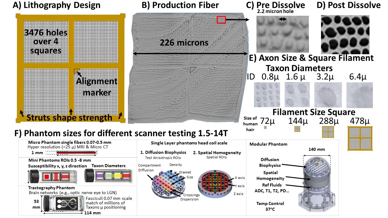
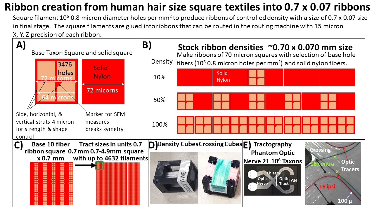
This project introduces to MRI phantom production bi-component polymer production control of Taxon shapes. In the 1990s the textile industry developed “logo textiles” with 0.45 micron pixel control of points to produce a filament of 50 microns. These are used in the apparel industry to create logo fibers used to catch counterfeiting of licensed apparel. Figure 1A shows the bi-component production. Two high pressure extrusion screws pump liquid polymers; one that will solidify (Nylon) and second that will dissolve in water Polyvinyl Alcohol (PVOH) ([WS1] . The Nylon is hydrophilic and water permeable, which allows water to enter the tubes, either from the end or through the wall over a 24 hour period, dissolving out the PVOH and making axon scale water tubes. The location and size of holes is based on lithography plates that provide .45 micron holes to define is the placement of the solid Nylon and dissolvable polymer. It uses polymer stretching to reduce the effective size of the fiber and pixels to an operating size of 0.45 micron [WS2] pixel control. This involves the spraying of thousands of high pressure jets of PVOH into Nylon polymer seas that are liquid and fall by gravity several meters, cooling to a glass transition solid that is stretched, cooled and spooled at a high rate (e.g., 60 m per minute). The production method can place Nylon and water selectively in intra and extra Taxonal compartments to control the restricted and hindered water spaces, hole diameters, and hold densities (Figure 1B, C). We produce Taxon fasciculi in a scale of 72 to 478 microns to produce hole sizes from 0.8 to 6.4 microns in phantoms of size ranging from 1 to 140 mm diameter to be scanned on MRI scanners from 1.5T to 14T. The filaments are combined into ribbons 0.07 x 0.7 mm and ROIs up to 4.9 mm square (Figure 3). Ground truth NIST traceable non-MRI measurement is provided by optical, SEM microscope imaging, and Micro-CT measurement (Figure1B, 2B-E, 4A, D). Analytical balances measure the fiber before and after dissolve, reporting weight loss averaging 48% and providing an indication of the amount of hole volume; also seen in SEM images (Figure 2BCD). [WS1]Critical request. Had used the incorrect chemical name of EVOH when it should have been PVOH here and in Figure 1 [WS2]Was ?? in submissionRESULTS
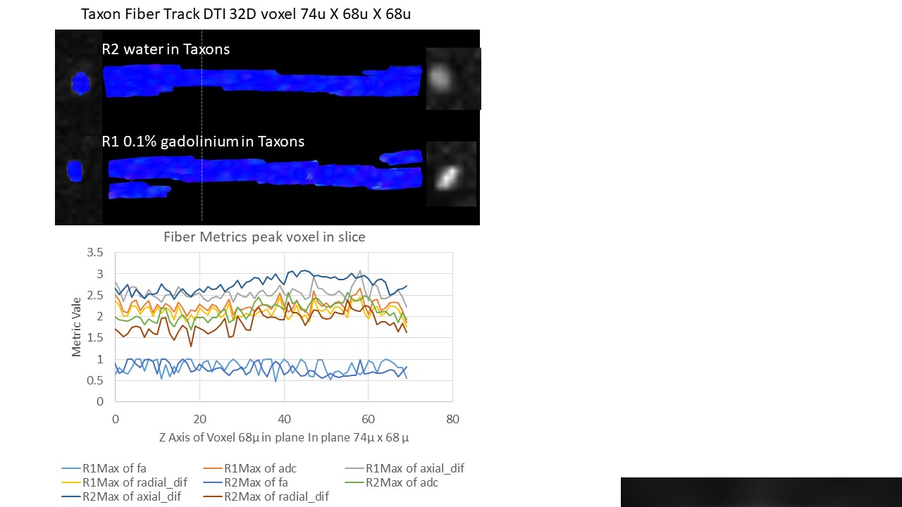
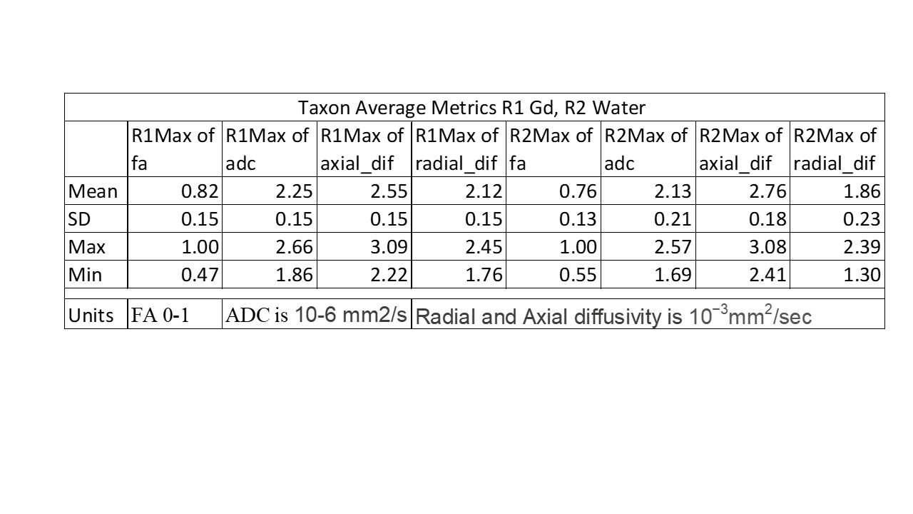
Taxon production methods can achieve human axonal scale of 0.8µm with 106 packing density per mm2 with a water fraction >0.45. With a 3x3 in plane voxel size and moving position we used the peak value that was for FA in the 1.0 range in Gd and mean 0.82, for water peak 1 and mean 0.62 (see Figure 4 & 5).DISCUSSION
These are early but encouraging results indicating we can provide ground truth measurement for precision of axonal diameters, water fractions, crossing angles, crossing densities, and Tractography-based point to point trajectories.CONCLUSION
The phantom can provide common calibrated measurement across laboratories with matched production reference measurement, advancing diffusion science and leading to quantitatively calibrated diffusion imaging.Acknowledgements
The novel textile phantom based on hollow fibers we developed with support from:
Contract Number: W911QY-15-C-0043; Title: Advanced Longitudinal Diffusion Imaging for TBI Diagnosis of Military Personnel
Contract Number: W81XH1220139; Title: High Definition Fiber Tracking Biological Diagnosis of TBI Providing Actionable Clinical Report of Quantified Damage
Award No. W81XWH-13-2-0095; Title: Chronic Effects of Neurotrauma Consortium
Award No: NIH U01EB026996
References
1. Innocenti, G.M., A. Vercelli, and R. Caminiti, The Diameter of Cortical Axons Depends Both on the Area of Origin and Target. Cerebral Cortex, 2014. 24(8): p. 2178-2188.
2. Caruyer, E., et al. Phantomas: a flexible software library to simulate diffusion MR phantoms. in ISMRM. 2014.
3. Chen, B. and A.W. Song, Diffusion tensor imaging fiber tracking with local tissue property sensitivity: phantom and in vivo validation. Magn Reson Imaging, 2008. 26(1): p. 103-8.
4. Chen, C.C., et al., Quality assurance of clinical MRI scanners using ACR MRI phantom: Preliminary results. Journal of Digital Imaging, 2004. 17(4): p. 279-284.
5. D'Souza, W.D., et al., Tissue mimicking materials for a multi-imaging modality prostate phantom. Medical physics, 2001. 28: p. 688-700.
6. Huber, J.S., Q. Peng, and W.W. Moses, Multi-modality phantom development. IEEE Transactions on Nuclear Science, 2009. 56: p. 2722-2727.
7. Komlosh, M.E., et al., Pore diameter mapping using double pulsed-field gradient MRI and its validation using a novel glass capillary array phantom. Journal of Magnetic Resonance, 2011. 208(1): p. 128-135.
8. Neher, P.F., et al., Fiberfox: facilitating the creation of realistic white matter software phantoms. Magnetic Resonance in Medicine, 2014. 72(5): p. 1460-1470.
9. Perrone, D., et al., D-BRAIN: anatomically accurate simulated diffusion MRI brain data. Plos One, 2016. 11(3): p. e0149778.
10. Yanasak, N. and J. Allison, Use of capillaries in the construction of an MRI phantom for the assessment of diffusion tensor imaging: demonstration of performance. Magnetic Resonance Imaging, 2006. 24: p. 1349-1361.
11. Matsuya, R., et al., A new phantom using polyethylene glycol as an apparent diffusion coefficient standard for MR imaging. International Journal of Oncology, 2009. 35: p. 893-900.
12. Lee, S., et al., Electrical conductivity estimation from diffusion tensor and T2: a silk yarn phantom study. Proceedings 14th Scientific Meeting, International Society for Magnetic Resonance in Medicine, 2006. 26: p. 3034.
13. Horkay, F., C. Pierpaoli, and P.J. Basser. Phantom for diffusion mri imaging. 2010 Januar1 26. 14. Ogrezeanu, G. and A. Hartlep. Fiber tracking phantom. 2009 Apri1 21.
15. Grech-Sollars, M., et al., Stability and reproducibility of co-electrospun brain-mimicking phantoms for quality assurance of diffusion MRI sequences. Neuroimage, 2018.
Figures
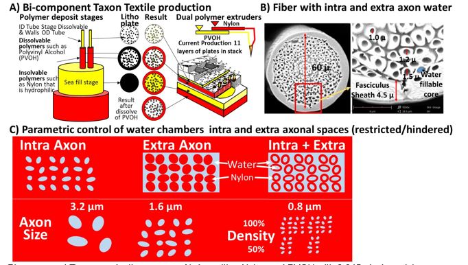
Bi-component Taxon production process A) depositing Nylon and PVOH with 0.045 pixel precision control of intra and extra Taxonal water compartments, B) producing Taxon water tubes with outside rings, C) with parametric control of size, density and position.