3061
Cerebral venous oxygen saturation variations in Alzheimer's disease complicated with type 2 diabetes patients using susceptibility weighted imaging mapping1The First Affiliated Hospital of Dalian Medical University, Dalian, China, 2GE healthcare, Beijing, China
Synopsis
Alzheimer's disease (AD) is a progressive neurodegenerative disease. Epidemiological studies suggest that type 2 diabetes (T2DM) patients are 2 times more likely to develop AD than healthy people. But it is unclear yet why more decreased saturation of blood oxygen and secondary more severe cognitive impairment are present in AD patients with diabetes. Susceptibility weighted imaging (SWI) is widely used in the diagnosis of central nervous system diseases and venous oxygen content is the basis of SWI angiography. As such, this study used SWI mapping to measure the changes of magnetic susceptibility as well as the change of blood oxygen.
Objective
To evaluate the change of cerebral venous oxygen saturation of Alzheimer's disease (AD) patients without T2DM (AD-DM) and with T2DM (AD+DM), and to explore the relevance between venous oxygen saturation and cognitive impairment, based on the quantitative analysis of cerebral venous oxygen saturation by using susceptibility weighted imaging mapping (SWIM).Materials and Methods
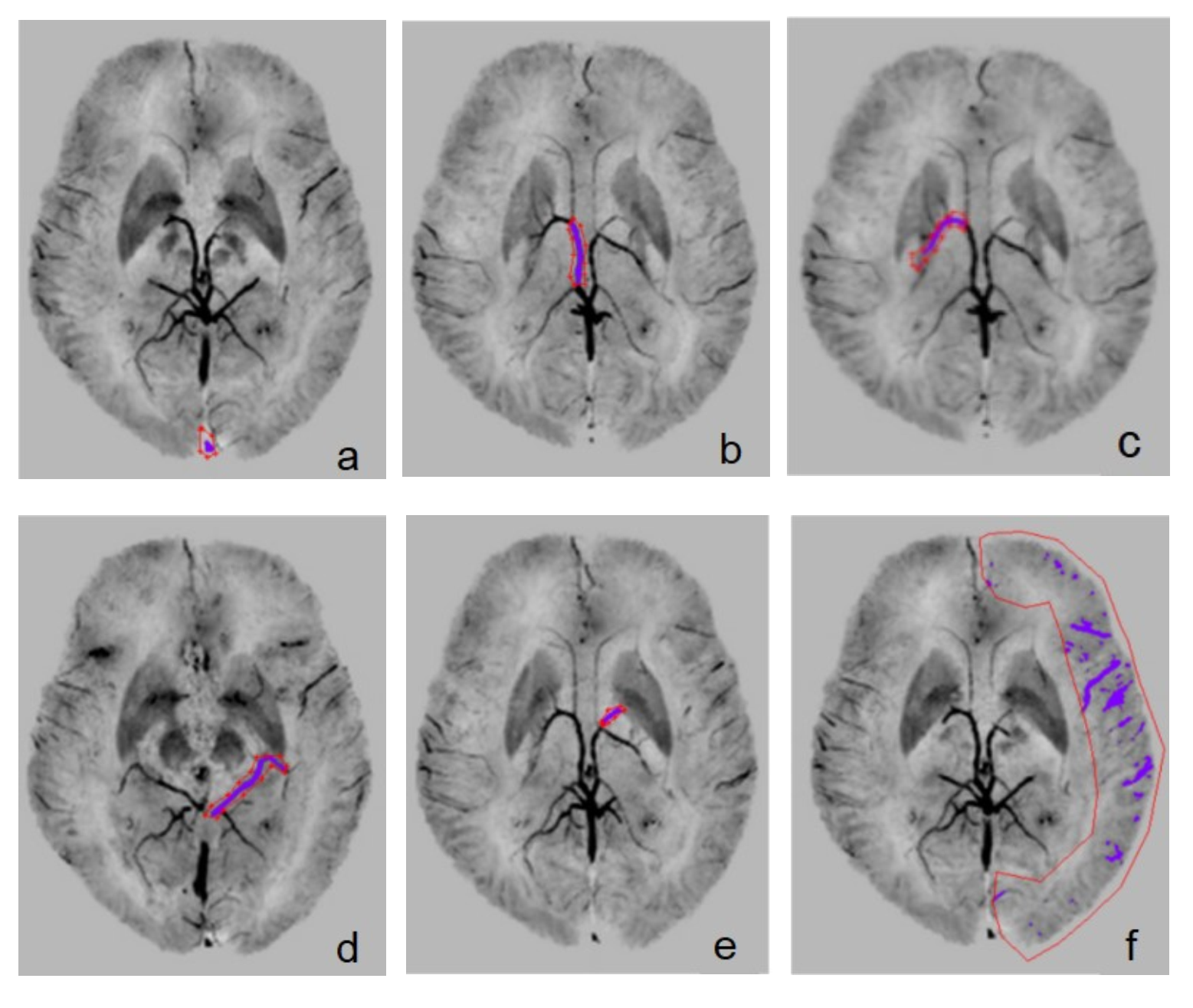
29 clinically confirmed AD-DM patients (7 males and 22 females, mean age = 72.86±9.29 yrs) and 12 AD+DM patients (4 males and 8 females, mean age = 75.50±7.36 yrs) were enrolled in this study. The cognitive scores (MMSE, MoCA, clock drawing test and activity of daily living (ADL)) and laboratory index (Fasting blood glucose, Triglyceride and total cholesterol) were collected. All subjects underwent routine MRI sequence and enhanced gradient echo T2* weighted angiography (ESWAN) on a 3.0T scanner. SWIM images were post-processed with signal processing in neuro-MR. The region of interests (ROIs) of bilateral cerebral veins (Superior sagittal sinus (SSS), cerebral cortical veins (CCV), venae thalamostriate (VTS), internal cerebral veins (ICV), basal veins of rosenthal (BV) and anterior caudate vein (ACV)) in SWIM were outlined manually and magnetic sensitivity value (MSV) was measured (Figure 1). For the values conformed to normal distribution, student t test was used; otherwise, the rank sum test was used. Spearman correlation was analyzed between the cognitive scores, laboratory index and MSV.Results
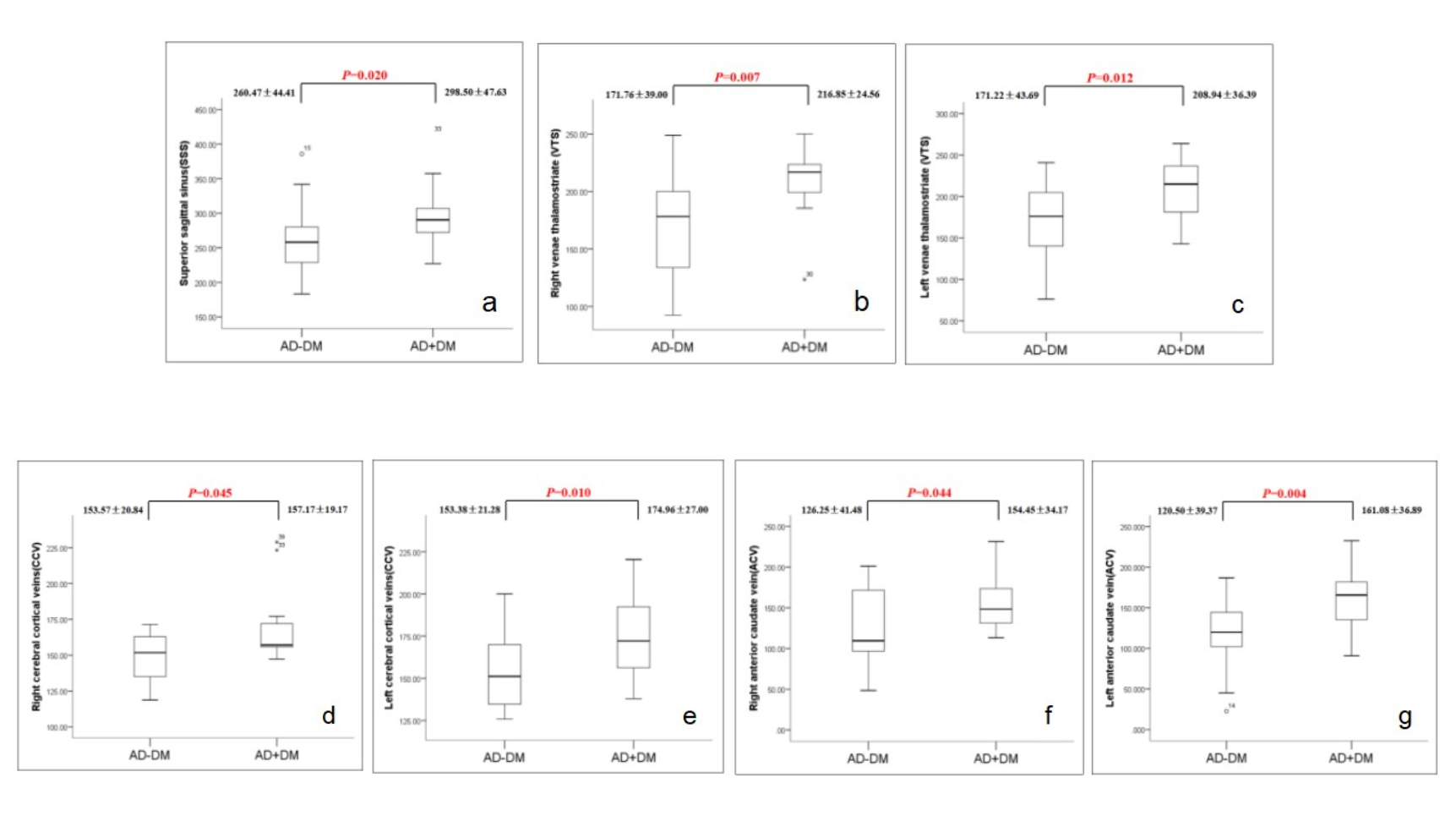
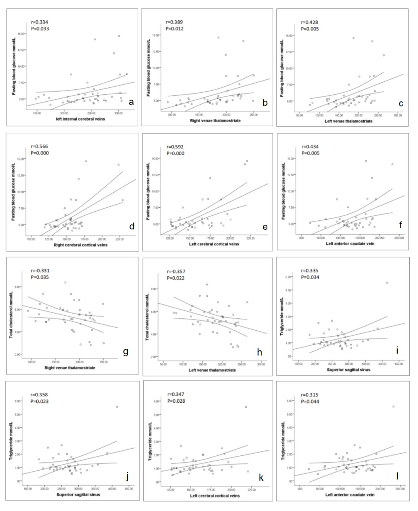
Compared to those of AD-DM, MSV of veins of AD+DM patients was increased, especially for that in SSS, bilateral VTS, CCV and ACV (Figure 2). There was no significant correlation between MSV and cognitive score. Fasting blood glucose was associated with left ICV, left ACV, bilateral VTS and CCV. Bilateral VTS was relevant to total cholesterol. Triglyceride was associated with SSS, bilateral CCV and left ACV (All P values<0.05) (Figure 3).Discussion
Venous oxygen saturation is an important indicator of brain function assessment[1]. This study used SWIM to measure the MSV and further the change of blood oxygen according toΔχvein-tissue=Κ×Δχdo×Hct(1-Y) (Δχ, Magnetic sensitivity; Y, Saturation of blood oxygen). The results indicate the blood oxygen level of AD+DM patients is lower than that of AD-DM patients. Cerebral small vessel diseases are common in diabetics. The pathological changes include the decrease of the number of capillaries, the increase of the permeability of the cerebral vessels and the thickening of the basement membrane of the capillary vessels[2-3]. This may be the cause of lower blood oxygen level in AD+DM. Compared to those of AD-DM patients, the bilateral cortical veins, thalamic striatum veins and anterior caudate nucleus veins of AD+DM showed more significant hypoxia, suggesting that cortical and basal ganglia vessels may be the earliest indicators. Fasting blood glucose, total cholesterol and triglyceride were significantly correlated with MSV, indicating that blood glucose and blood lipid were influencing factors of blood oxygen. No correlation between MSV and cognitive score was found, maybe because cognition affected by the change of blood oxygen is a long evolutionary process.Conclusion
Cerebral venous oxygen saturation was decreased in patients of AD complicated with DM than AD without DM, which correlated with hyperglycemia and hyperlipidemia.Acknowledgements
No acknowledgement found.References
[1]van Norden AG, van Dijk EJ, de Laat KF, et al. Dementia: Alzheimer pathology and vascular factors: From mutually exclusive to interaction. Biochim Biophys Acta ,2012,1822(3):340-349.
[2] Fan AP, Benner T, Bolar DS, et al. Phase-based regional oxygen metabolism (PROM) using MRI[J]. Magn Reson Med, 2012, 67(3): 669-678.
[3] Dalal PM, Parab PV. Cerebrovascular disease in type 2 diabetes mellitus. NeurolIndia, 2002, 50(4): 380-385.
Figures