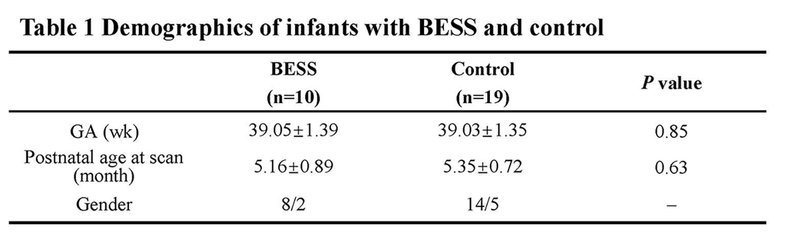
3026
Alterations of brain white matter in infants with begin enlargement of subarachnoid space by assessing conventional diffusion and WMTI metrics.1Department of Radiology, Department of Radiology, The First Affiliated Hospital of Xi’an Jiaotong University, Xi'an, China
Synopsis
BESS is characterized by increased cerebrospinal fluid in subarachnoid spaces; some infants with BESS were accompanied with mildly motor and language delay. White matter (WM) development is important to neurodevelopmental outcomes, but relationships between BESS and WM maturation are not very clear. This study aims to quantitative assess WM microstructures of in infant with BESS aged 4-6 months by conventional diffusion and WMTI metrics. Significant decreased FA, α and increased RD, AD, MD, RDe were found in infants with BESS. All results suggested underlying alterations of WM, especially for myelination outside axons. It may provide additional information for neurodevelopment outcomes.
Introduction
Begin enlargement of the subarachnoid space (BESS) is characterized radiologically by increased cerebrospinal fluid in the subarachnoid spaces, usually bifrontal, with a widened interhemispheric fissure and normal to slightly increased ventricular size[1]. Previous studies found that some children with BESS were accompanied with mildly motor and language delay. However, relationships between the enlargement of subarachnoid space and brain development are not very clear. Since white matter (WM) development plays an important role in neurodevelopmental outcomes of children, the quantitative measurements may find more subtle alterations to assess the brain maturation. But there are few studies investigating WM development of children with BESS. This study aims to compare WM microstructures of a pediatric population aged 4-6 months with BESS to controls using conventional diffusion metrics and white matter tract integrity (WMTI) metrics from diffusion kurtosis imaging (DKI).Materials and Methods
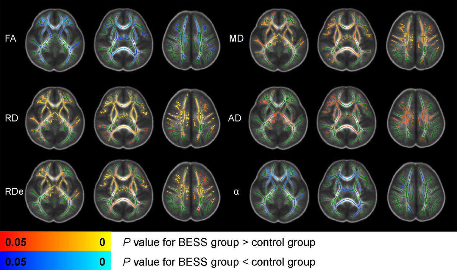
The Institutional Review Broad of the first author’s affiliation approved this study and written informed consents were obtained from parents of the children. Patients For our study, BESS were defined as a frontal subarachnoid space or interhemispheric fissure greater than 5mm on transverse T2-weighted imaging[2,3]. Infants with BESS and controls aged 4-6 months were included in the study. Subjects with abnormalities of brain parenchyma on MRI were excluded. All these infants completed scan of conventional and DKI sequences. MR Protocols All subjects were examined by using a 3.0T scanner (Signa HDxt, General Electric Medical System, Milwaukee, WI, USA) with an 8-channel head coil. Data acquisition included three-dimensional fast spoiled gradient-echo T1-weighted sequence (TR/TE, 10.2ms/4.6ms; NEX, 1; isotropic 1×1×1mm3; FOV, 24cm) and transverse fast spin-echo T2-weighted sequence (TR/TE, 4200ms/116ms; NEX of 1.5; matrix, 320×320; thickness, 4mm; FOV, 24cm), followed by a DKI (25 directions; b value, 500, 1000, 1500, 2000, 2500 s/mm2; SENSE factor, 2; TR/TE, 11000/93.9ms, slice thickness, 4mm with 4mm gap, FOV, 24cm; matrix, 172×172). Data and statistical analysis DKI raw data were preprocessed by FMRIB software library (FSL; http://www.fmrib.ox.ac.uk/fsl) and four parameters of fractional anisotropy (FA), mean diffusivity (MD), axial diffusivity (AD), and radial diffusivity (RD) and WMTI metrics of intra-axonal axial diffusivity (ADa), intra-axonal radial diffusivity (RDa), extra-axonal axial diffusivity (ADe), extra-axonal radial diffusivity (RDe), tortuosity (α) were calculated by using the FMRIB Diffusion Toolbox (http://fsl.fmrib.ox.ac.uk/fsl/fslwiki/FDT). Here we use the local FA map as a reference. Wilcoxon signed-rank tests were used for the differences in demographics between two groups. Conventional diffusion metrics and WMTI metrics of the white matter were calculated by general linear model. p<0.05 was considered as statistically significant difference.Results
A total of 10 infants with BESS and 19 controls at age of 4-6 months were enrolled. No significant differences in GA, postnatal age at MRI scan were found between groups (Table 1). Compared with controls, significantly decreased FA, increased RD, AD, MD were observed in infants with BESS group. For WMTI metrics, increased RDe and decreased α were also founded (Figure 1). The distribution of significance results were mainly in the anterior regions of brain, such as genu of corpus callosum, internal capsule and anterior corona radiata.Discussion
Enlargement of the subarachnoid space is a specific radiographic pattern, yet its pathogenesis has not been established. It may be related to arachnoid villi maturity or different growth rates of brain and skull in infants[4]. Previous study suggested FA was increased in BESS due to the compression of brain[5]. Our results also suggested the BESS has relationship with alterations of WM, but the changes of diffusion metrics were contrary to their study. Decreased RD and RDe could potentially reflect the myelination of WM, especially RDe reflected the outside structure of axons. Our results suggested RD and RDe of children with BESS was higher than controls and FA was decreased in infants with BESS. It may indicated the maturation of myelination in infants with BESS was delay. Further we will follow-up neurodevelopment outcome of these infants with BESS to further validate the relationships with alterations of WM.Conclusions
BESS may affect the white matter development at age of 4-6 months, especially for the myelination outside the axons. It may provide additional information for the neurodevelopment outcomes.Acknowledgements
No acknowledgement found.References
1. Zahl SM, Egge A, Helseth E, Wester K (2011) Benign external hydrocephalus: a review, with emphasis on management. Neurosurg Rev 34 (4):417-432. doi:10.1007/s10143-011-0327-4 2.
2. Nickel RE, Gallenstein JS (1987) Developmental prognosis for infants with benign enlargement of the subarachnoid spaces. Developmental medicine and child neurology 29 (2):181-186
3. Prassopoulos P, Cavouras D, Golfinopoulos S, Nezi M. The size of the intra- and extraventricular cerebrospinal fluid compartments in children with idiopathic benign widening of the frontal subarachnoid space. Neuroradiology 1995;37:418-21
4. Paciorkowski AR, Greenstein RM (2007) When is enlargement of the subarachnoid spaces not benign? A genetic perspective. Pediatr Neurol 37 (1):1-7. doi:10.1016/j.pediatrneurol.2007.04.001
5. Sun M, Yuan W, Hertzler DA, Cancelliere A, Altaye M, Mangano FT (2012) Diffusion tensor imaging findings in young children with benign external hydrocephalus differ from the normal population. Child's nervous system : ChNS : official journal of the International Society for Pediatric Neurosurgery 28 (2):199-208. doi:10.1007/s00381-011-1651-2
Figures