2435
Efficient Motion-Corrected, Model-Driven Reconstruction for Simultaneous Multi-Contrast MPnRAGE1University of Wisconsin, Madison, WI, United States
Synopsis
A model-based denoising algorithm is developed for efficient reconstruction of a large number of images with different T1 contrasts using MPnRAGE. The method takes only 50% the time of a single iteration of traditional constrained reconstruction methods.
Introduction
MPnRAGE [1] is a technique to generate a large number of high resolution, 3D MPRAGE images with different T1-contrasts (i.e. different TIs or inversion times) as well as a quantitative T1 map from a single scan that is about the same duration as a fully sampled MPRAGE acquisition. It utilizes 3D radial k-space sampling, which makes it less sensitive to motion (blurring as opposed to ghosting) and is amenable to retrospective motion correction [2].
In the original implementation, a sliding window k-space reconstruction used a tornado [3] or KWIC [4] filter for larger view-sharing of higher spatial frequency components than low-spatial frequency components. This created a tremendous computation burden as roughly 50% of the total (non-Cartesian) samples underwent a gridding operation for each of the approximately 400 frames.
An alternative option eliminates view-sharing, so that each frame only uses a small portion of the data (~1/400th). However, the resulting images are severely undersampled (factor ~500 to 1000), which presents serious challenges for compressed sensing techniques. Those techniques would also require many iterations (50-200 typically), which would possibly be slower than the KWIC filter due to additional time from several forward and backwards FFTs during each iteration, let along the gridding operations.
Here, we propose a model-based denoising technique that is applied to images formed without view-sharing or iterative compressed sensing techniques, and thus, provides a fast and effective way to provide high quality T1-weighted images.
Methods
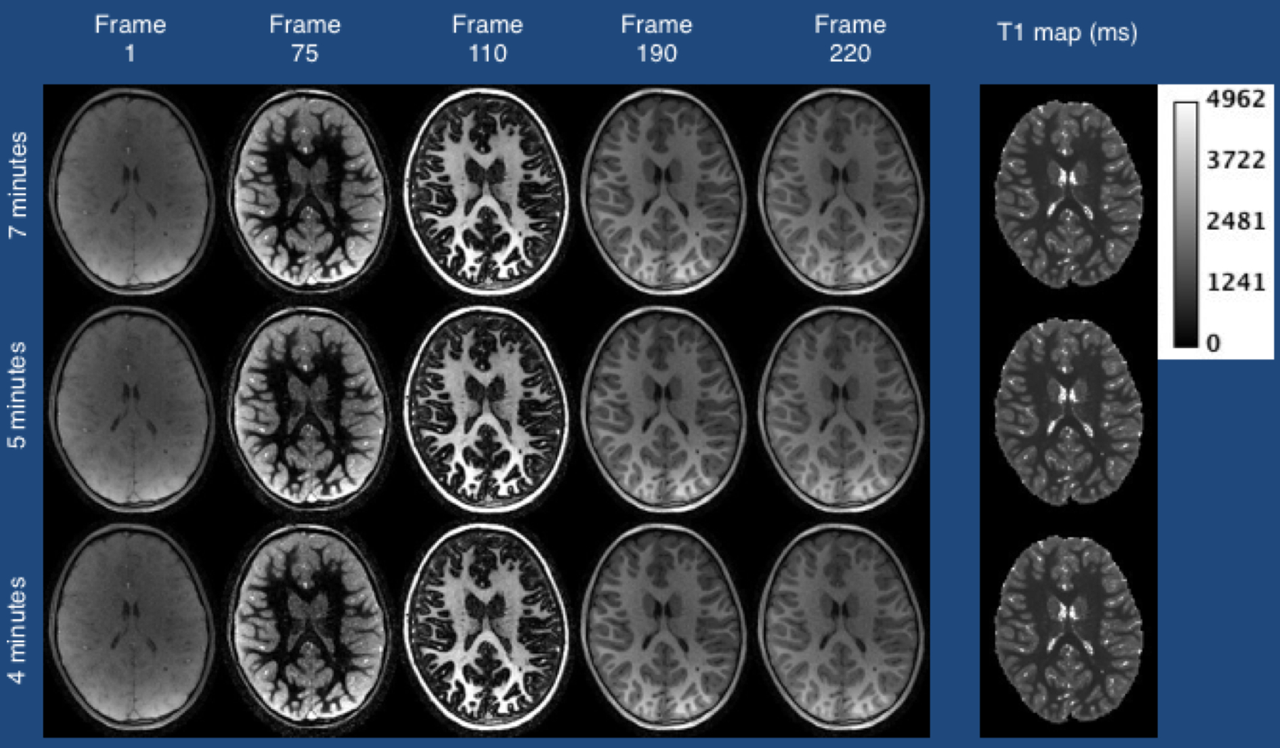
A 6 year old boy was scanned on a 3T MR750 (GE Healthcare) scanner without sedation. 3D images with whole brain coverage and 1.0 mm isotropic resolution were acquired using a modified MPnRAGE sequence similar to [1]. Parameters included delay time TD=500 ms, TR=4.9 ms, TE=1.8 ms,n=386 views along the recovery curve starting with a minimum TI of 15 ms. The excitation flip angles were 4°/8°for the first 325/remaining 61 views. The scan time was 7 minutes, although processing (below) was also performed using only the first 4 and 5 minutes of data to observe differences in image quality with shorter scan durations.
Prior to parametric fitting, rotational and translation motion was corrected for according to [2].
Images from each point along the recovery curve were reconstructed without parallel imaging, compressed sensing, or view-sharing from neighboring points along the recovery curve. A two pass fitting procedure first fit for all four unknown parameters (T1, spin-density, B1, and inversion efficiency). The resulting B1 and inversion efficiency images were then smoothed (gaussian filters FWHM of 7mm and 9mm) and treated as known parameters for a second fitting of the original data to determine T1 and spin-density. The resultant T1 and spin-density maps were denoised with TV minimization using a small regularization parameter (optional step). The final maps were used to regenerate the signal for each voxel at each of the measured points along the inversion recovery curve, thereby providing a “denoising” of the original images.
Results
Examples of the raw and denoised images for a 4 minute scan (undersampled by over 1000) are shown in Fig. 1. Example images demonstrating five points along the inversion recovery curve, as well as the T1 map, are show in Fig. 2 both with and without motion correction. Motion creates a uniform blurring that does not impede the model-based denoising algorithm and is effectively reduced with motion correction. Figure 3 shows motion corrected images from five points along the inversion recovery curve when subsets of the data are used. The results suggest that there is little loss of image quality using only a 4 minute scan.Discussion
The method presented in this work utilizes only a single gridding and FFT operation for each inversion frame, and thus, takes only about only ¼ to ½ the time of a single iteration of gradient based methods (with or w/o compressed sensing regularization terms). It is therefore likely 50-200 times faster than compressed sensing options, and is about 50 times faster than the view-sharing method originally proposed. The total computation time is about 1 hour (50 minutes for image reconstruction and 10 for denoising) using a 24-core machine with Intel Zeon 2.6 GHz processors.Conclusions
An efficient, model-based denoising technique was introduced that allowed a large number of differently T1-contrasted MPnRAGE images to be produced without the need of computationally expensive algorithms.Acknowledgements
No acknowledgement found.References
1. Kecskemeti S, et al., MPnRAGE: A technique to simultaneous acquire hundreds of differently contrasted MPRAGE images with applications to quantitative T1 mapping. Magn Reson Med. 75:1040-1053 (2016)
2. Kecskemeti S, et al., Robust Motion Correction Strategy for Structural MRI in Unsedated Children Demonstrated with Three-dimensional Radial MPnRAGE. Radiology 2018; 289:509-516
3. Barger A, et al. Time-Resolved Contrast-Enhanced Imaging With Isotropic Resolution and Broad Coverage Using an Undersampled 3D Projection Trajectory. Magn Reson Med. 48:297-305 (2002)
4. Song H, et al. k-Space Weighted Image Contrast (KWIC) for Contrast Manipulation in Projection Reconstruction MRI. Magn Reson Med. 44:825-832 (2000)
Figures
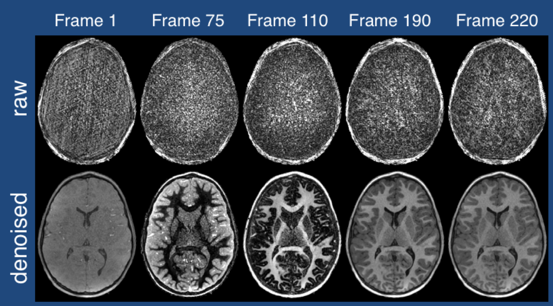
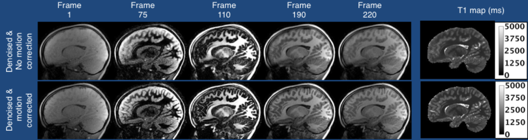
Figure 2: Select T1-contrasts and T1 maps (in ms) shown without (top) and with (bottom) retrospective motion correction. Note that the non-motion corrected images are blurry, but motion correction has effectively reduced the blurring. In all cases, the model based denoising has been performed, which is not negatively affected by either motion artifacts (blurring) or the addition of motion correction.