2051
Clinical Performance of Non-contrast Whole-heart Magnetic Resonance Coronary Angiography with Compressed Sensing: Comparison with Conventional Sensitivity Encoding ImagingYuki Ohmoto-Sekine1, Junji Takahashi2, Norihito Miura2, Saori Amemiya2, Kei Fukuzawa2, Makiko Ishihara3, Hiroshi Tsuji1, and Yasuji Arase1
1Health Management Center, Toranomon Hospital, Tokyo, Japan, 2Radiology Dept., Toranomon Hospital, Tokyo, Japan, 3Imaging Center, Toranomon Hospital, Tokyo, Japan
Synopsis
Non-contrast whole-heart magnetic resonance coronary angiography (WHMRA) has proven value for noninvasive assessment of coronary arteries. However, there are major technical challenges associated with this technique, such as image quality degradation due to long scan times. Compressed sensing (CS) reconstruction might be a powerful solution for shortening scan time. We conducted a feasibility study for WHMRA with conventional parallel imaging and CS reconstruction. WHMRA with CS reconstruction is a promising method that can shorten the scan time while maintaining mostly acceptable images, although there is still room for improvement, especially for stenotic vessel evaluation.
Introduction and Purpose
Non-contrast whole-heart magnetic resonance coronary angiography (WHMRA) [1] is very useful and safe in screening for coronary artery disease. However, there are major technical challenges associated with this technique, such as image quality degradation due to respiratory motion and long scan times. Compressed sensing [2] reconstruction of under-sampled measurements generates missing data based on assumptions of image sparsity. A short scan time derived from compressed sensing is beneficial for performing WHMRA. This study aimed to determine whether WHMRA with compressed SENSE (CS-SENSE) reconstruction can take the place of WHMRA with conventional sense encoding (SENSE) reconstruction in clinical settings by health checkups.Methods
With institutional review board approval and informed consent, 51 subjects underwent MR imaging on a commercial 1.5-T scanner (Ingenia, Philips Healthcare) with a digital broadband system, using a phased-array torso coil (dS Torso coil). Electrocardiogram-triggered, navigator-gated, magnetization-prepared (T2-prep, fat-suppressed), 3D-balanced steady-state free precession (bSSFP) sequence was performed with the following parameters: TR/TE/FA = 3.2 ms/1.6 ms/70° and spatial resolution = 1.6 × 1.6 × 1.6 mm3 interpolated to 0.8 × 0.8 × 0.8 mm3. WHMRA was performed with (i) a conventional approach using SENSE reconstruction with factors of 2.3 in phase and 1.4 in slice directions, and (ii) a factor of 5 CS-SENSE reconstruction. We compared the scan times, image quality, visible vessel length, vessel sharpness, vessel diameter, and vessel contrast. The WHMRA data were transferred to a workstation (ZIO, Amin Ltd., Tokyo, Japan) to make curved multiplanar reformation and the image quality was assessed using a 5-point scale (1, poor/nondiagnostic; 2, fair/moderate blurring; 3, acceptable/mild blurring; 4, good/no blurring; and 5, excellent/sharp borders) by an experienced observer using randomized image pairs. A Wilcoxon signed-rank test was used to compare image quality, and a paired t-test was used to assess the scan times, vessel length, vessel sharpness, vessel diameter, and vessel contrast.Results and Discussion
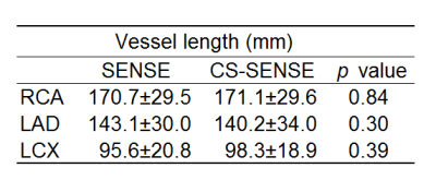
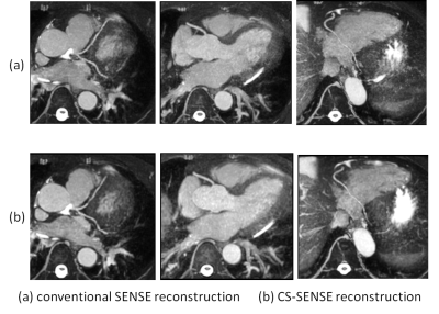
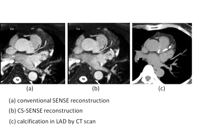
The mean effective scan time was significantly reduced with CS-SENSE reconstruction (7 min 01 s for SENSE vs. 4 min 47 s for CS-SENSE p< 0.001). All quality and quantity evaluations of CS-SENSE reconstruction were acceptable. Vessel length (Table 1) (right coronary artery [RCA] SENSE vs. CS-SENSE p= 0.84, left anterior descending [LAD] SENSE vs. CS-SENSE p= 0.30, and left circumflex [LCX] SENSE vs. CS-SENSE p= 0.39) was not significantly different. Image quality was not significantly different (Table 2). Vessel diameter showed no significant difference between two reconstructions, vessel contrast in each segment of the RCA and proximal LCX with CS-SENSE reconstruction was lower, and vessel sharpness in the distal RCA and mid LAD with CS-SENSE reconstruction was lower compared to that with SENSE reconstruction (Table 3). Motion may lead to differences in vessel contrast results in each segment of the RCA, which has a shorter acquisition window and greater movement compared to the LAD and LCX. On the other hand, the differences in sharpness in the LAD might be due to low sparsity between the signal intensity of the coronary artery and surrounding myocardium. In some images with CS-SENSE reconstruction, poor image quality in the mid segment of the LAD was observed and signal loss was found in a calcified artery, even when the artery was patent (Fig2).Conclusion
WHMRA with CS-SENSE reconstruction is a promising method that can shorten the effective scan time while maintaining a mostly acceptable image, although there is still room for improvement, especially for stenotic vessel evaluation. Further development is warranted for CS-SENSE reconstruction.Acknowledgements
No acknowledgement found.References
[1] Weber OM, Martin AJ, Higgins CB. Magn Reson Med. 2003; 50:1223-8. [2] Lustig M, Donoho D, Pauly JM. Magn Reson Med. 2007; 58:1182-95.Figures

table1. Visible
vessel length measurement between SENSE reconstruction and CS-SENSE reconstruction.

table 2. Image scores between SENSE reconstruction and CS-SENSE reconstruction. prox, proximal segment; mid, mid segment; dis, distal segment.

Table3. Qualititative analysis between SENSE reconstruction and CS-SENSE reconstruction. pro, proximal segment; mid, mid segment; dis, distal segment.

Fig 1. Typical reformatted image obtained using (a) conventional SENSE
reconstruction and (b) CS-SENSE reconstruction.

Fig2. Example from one
representative image obtained using (a) conventional SENSE reconstruction and
(b) CS-SENSE reconstruction. Signal loss was observed (arrow) in the mid LAD (b) with
calcification (c), even when the artery was patent (a).