1480
Multi-Coil Array for Combined Imaging and B0 Shimming in a Portable Head-Only Scanner1Department of Biomedical Engineering, Columbia University in the City of New York, New York, NY, United States, 2Center for Magnetic Resonance Research, Department of Radiology, University of Minnesota, Minneapolis, MN, United States, 3Robinson Research Institute, Victoria University of Wellington, Wellington, New Zealand, 4Department of Radiology, Columbia University in the City of New York, New York, NY, United States
Synopsis
To improve accessibility of MRI and to enable advanced studies on motor coordination, we are part of the U01EB025153 collaboration to develop a compact 1.5 T head-only scanner enabling free limb movement. The compact size of the magnet significantly increases B0 inhomogeneity, necessitating the use of novel imaging concepts robust to B0 inhomogeneities that in turn require advanced B0 field modeling capabilities. Here, we introduce a 31-coil Multi-Coil (MC) design capable to generate linear and non-linear MR image encoding fields as well as complex B0 shim fields with low space and power requirements.
Introduction
While MRI offers unique capabilities to visualize human brain anatomy and conduct neurobehavioral studies, its accessibility is limited to large institutions due to the significant costs and infrastructural requirements of MR scanners. Furthermore, subjects must lie supine during measurement. To address these issues, we are part of a collaboration (NIH U01EB025153) to build a novel compact 1.5 T head-only scanner. This new design allows free limb movement and subject vision through a viewing window to enable novel research on motor coordination and social interaction.
The small size of the magnet, however, inevitably leads to significant B0 imperfections that exceed the limits of standard MR image encoding. We therefore apply recently introduced concepts with a high tolerance to B0 inhomogeneity like missing-pulse steady-state free precession1,2 (MP-SSFP) and STEREO3,4 with both linear and non-linear encoding. The high flexibility of the dynamic multi-coil (MC) technique, or DYNAMITE, is uniquely suited to generate linear5,6 and non-linear7 imaging fields along with concomitant complex B0 shim fields8 with a compact setup while maintaining a low power demand, thereby fitting a portable and compact scanner. Here, we present the mechanical design of a suitable MC configuration and theoretically assess its DYNAMITE field generation capabilities.
Methods
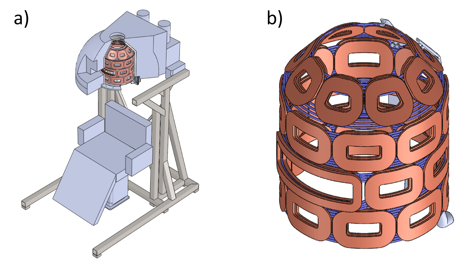
The MC system was designed in Solidworks (Dassault Systèmes, Vélizy-Villacoublay, France) based on the geometric constraints of the head-only scanner. It consists of 31 coils, with 200 turns each of copper wire, that will be individually driven with a maximum current of 5 A. Matching the geometry of the magnet bore, these coils are arranged in four rings of eight coils each with the lower three rings forming a cylinder and a tapered top ring (Figure 1). To incorporate the scanner’s window two coils in the second ring are fused into a bigger one. The whole system will be encapsulated in epoxy resin to increase mechanical stability.
Since the system will be used to create imaging fields and for B0 shimming, MC fields will be applied continuously during sequence execution. In addition, due to the 1.5 kHz average inhomogeneity of the scanner’s B0 field, shimming is significantly more demanding than in traditional MRI systems, thereby requiring the dedication of a larger portion of the system’s dynamic range. This might result in a high duty cycle for the system. We expect individual coils (resistance 1.8 Ω) to produce up to 45 W Joule heating. The system will consequently be water-cooled and the temperature continuously monitored using resistance temperature detectors to ensure subject and operational safety.
To assess the field generation capabilities of this design, coil wire paths were exported from the CAD model to PUMCIN9 format using a custom Add-in for Solidworks10,11. These coil patterns formed the basis for Biot-Savart simulations in B0DETOX11,12 of MC-generated fields within an elliptical region of interest (ROI, diameter 15x20x15 cm3) covering a subject’s head. Maximum temperatures in individual coils were estimated for the currents at hand using a steady-state finite element thermal simulation in Solidworks.
Results
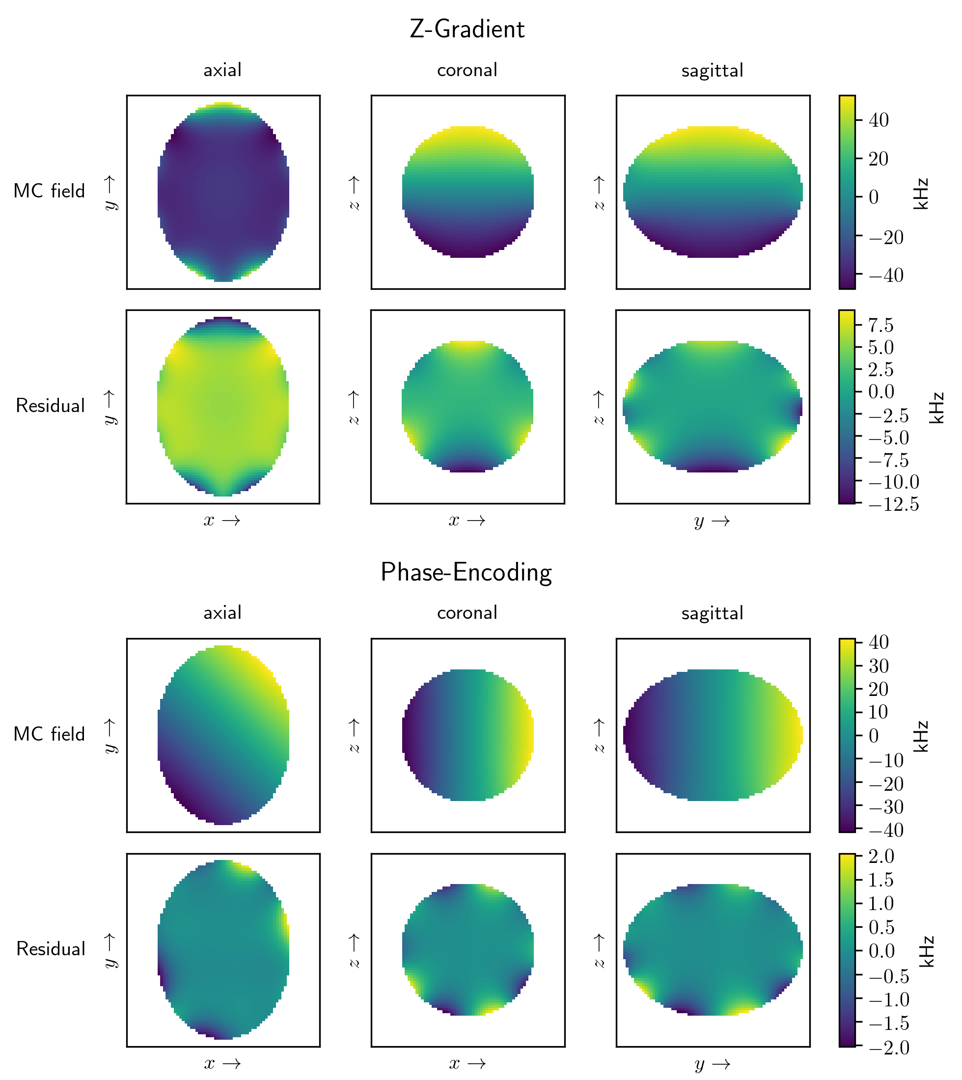
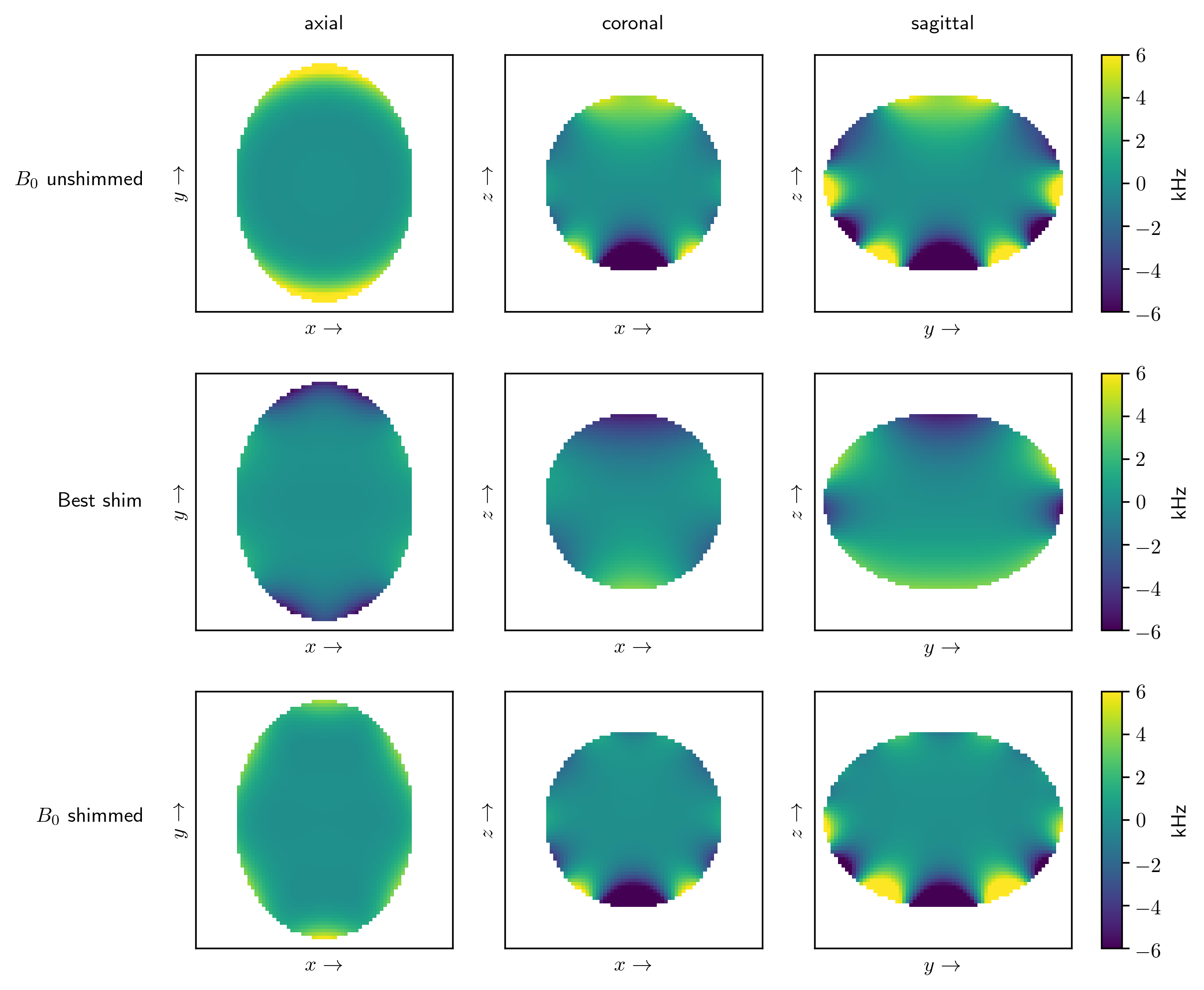
Linear encoding fields were generated as an 8 kHz/cm Z-gradient and as two orthogonal simultaneously applied 4.2 kHz/cm phase-encoding gradients in the axial plane (Figure 2). The generated fields had a median error of 783 Hz and 99 Hz, respectively. A simulated MC shim of the scanner’s B0 background field reduced the median field deviation from 726 Hz to 386 Hz, however, significant local inhomogeneities remained (Figure 3).
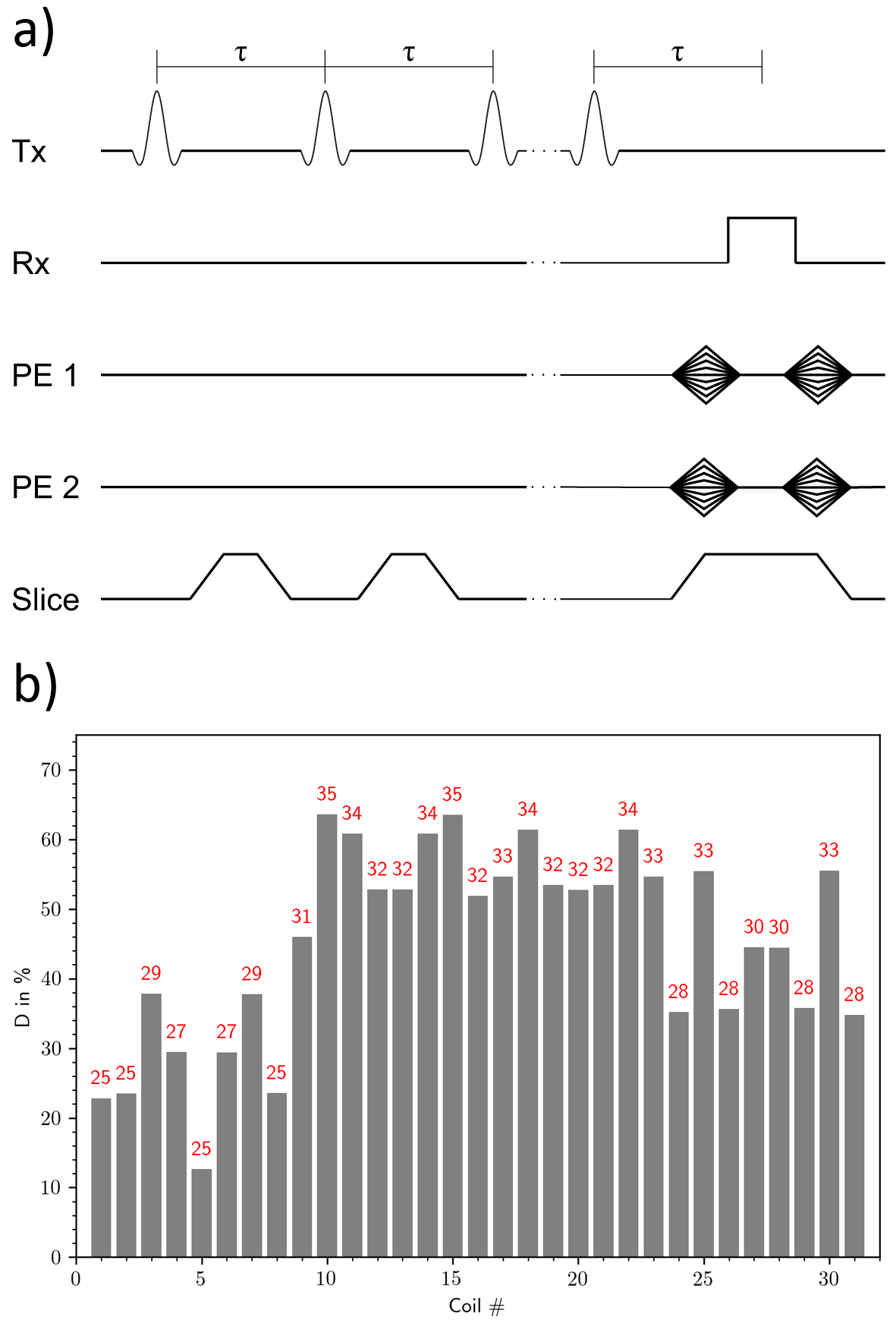
The duty cycle per coil for an MP-SSFP sequence (Figure 4a) was calculated based on simulated MC fields. Individual duty cycles ranged from 12.6 % to 63.6 % (average over entire setup: 51 %) and maximum temperatures in the coils did not exceed 35 °C (Figure 4b).
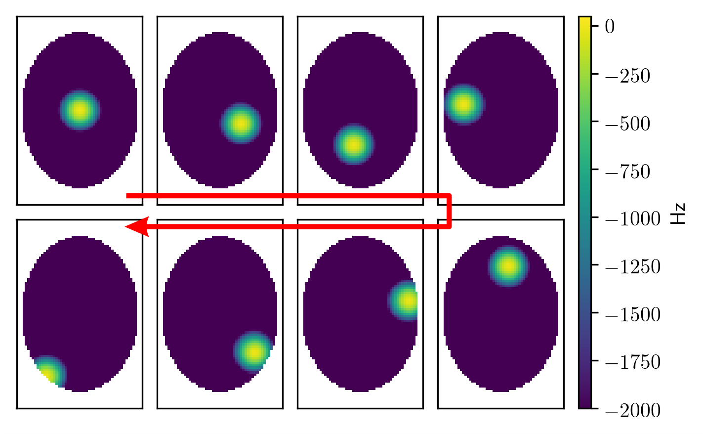
To assess non-linear field generation capabilities for STEREO, a 500 Hz/cm² parabolic focus was generated at different positions along a spiral trajectory throughout the elliptical ROI covering the human head (Figure 5). The median error of these individual field generations ranged from 2 Hz to 9 Hz.
Conclusion & Outlook
The presented simulations show that the proposed MC
setup is capable of generating linear and non-linear image encoding fields
strong enough for MR imaging throughout the extent of a human head and over the
entire execution time of an MRI sequence. Moreover, though this is not the
primary goal of the MC hardware, the setup is also capable of improving the uniformity
of the B0 field. The achieved uniformity is well within the capability of the novel imaging concepts to be used.
The next step comprises manufacturing the proposed MC hardware and subsequent experimental validation of the presented simulation results to enable advanced MR imaging with this novel compact head-only scanner.
Acknowledgements
This research was supported by the National Institute of Biomedical Imaging and Bioengineering of the National Institutes of Health under award number U01EB025153.References
1. Patz S, Wong STS, Roos MS. Missing pulse steady‐state free precession. Magn Reson Med. 1989;10(2):194-209.
2. Kobayashi N, Idiyatullin D, Adriany G, Garwood M. Magnetic Resonance Imaging under Highly Inhomogeneous B0 Fields using Missing-Pulse Steady-State Free Precession (MP-SSFP). In: Proc. Intl. Soc. Magn. Reson. Med. 25, 5052 (2017).
3. Snyder ALS, Corum CA, Moeller S, Powell NJ, Garwood M. MRI by steering resonance through space. Magn Reson Med. 2014;72(1):49-58.
4. Jayatilake ML, Juchem C, Mullen M, Adriany G, de Graaf RA, Garwood M. STEREO-MC for Connected Spatiotemporal Excitation. In: Proc. Intl. Soc. Magn. Reson. Med. 24, 2198 (2016).
5. Juchem C, Nixon TW, McIntyre S, Rothman DL, de Graaf RA. Magnetic field modeling with a set of individual localized coils. J Magn Reson. 2010;204(2):281-289.
6. Rudrapatna UA, Fluerenbrock F, Nixon TW, de Graaf RA, Juchem C. Combined imaging and shimming with the Dynamic Multi-Coil Technique. Magn Reson Med. 2018;0:1-10.
7. Juchem C, Nixon TW, de Graaf RA. Multi-Coil Imaging with Algebraic Reconstruction. In: Proc. Intl. Soc. Magn. Reson. Med. 20, 2545 (2012).
8. Juchem C, Nixon TW, McIntyre S, Boer VO, Rothman DL, de Graaf RA. Dynamic multi-coil shimming of the human brain at 7 T. J Magn Reson. 2011;212(2):280-288.
9. Juchem C, de Graaf RA. The public multi-coil information (PUMCIN) policy. Magn Reson Med. 2017;78(5):2042-2047.
10. Theilenberg S, Shang Y, Tirumala SS, Juchem C. Development of Multi-Coil B0 Technology with Computer-Aided Design Software. In: Proc. Intl. Soc. Magn. Reson. Med. 26, 4316 (2018).
11. MR Scientific Engineering for Clinical Excellence (MR SCIENCE) Laboratory software download. http://juchem.bme.columbia.edu/software-and-tools.
12. Juchem C, Herman P, Sanganahalli BG, Brown PB, Mcintyre S, Nixon TW, Green D, Hyder F, de Graaf RA. DYNAmic Multi-coIl TEchnique (DYNAMITE) shimming of the rat brain at 11.7T. NMR Biomed. 2014;27(8):897-906.
Figures