1261
Regional thigh muscle composition based on chemical shift encoding-based water-fat MRI and its association with muscle strength1Department of Neuroradiology, Klinikum rechts der Isar, Technical University of Munich, Munich, Germany, 2Department of Radiology, Klinikum rechts der Isar, Technical University of Munich, Munich, Germany, 3Department of Sport and Health Sciences, Klinikum rechts der Isar, Technical University of Munich, Munich, Germany
Synopsis
Chemical shift encoding-based water-fat MRI derived proton density fat fraction (PDFF) of the thigh muscles bears potential as a surrogate marker in subjects with osteoarthritis, sarcopenia, and neuromuscular disorders. Muscle PDFF has shown to correlate with isometric strength at the thigh and spine. However, MR-based muscle fat quantification requires time-consuming segmentation of multiple muscle compartments. Therefore, we investigated if segmentation of single compartment muscles and of different levels of the thigh influences the relation of PDFF to isometric strength. The present study demonstrated that PDFF measurements can be limited to an entire muscle compartment, independent of sampling level.
Introduction
Assessment of the thigh muscle fat composition by MRI has been proposed as a surrogate marker in subjects with osteoarthritis, sarcopenia, and neuromuscular disorders [1,2]. Chemical shift encoding-based water-fat MRI allows to reliably extract the proton density fat fraction (PDFF) of different muscle compartments [3]. First studies were able to relate intramuscular PDFF to strength measurements of quadriceps [4] and paraspinal muscles [5]. However, it is not known whether assessment of intra- or intermuscular fat should be favored or if the sampling level at the thigh plays a role. The purpose of this study was to investigate the association of isometric physical strength with intra- and intermuscular PDFF of the hamstring and quadriceps femoris muscles at different levels.Methods
Subjects:
Twenty-nine healthy subjects (15 women, 14 men; mean age 30.1 ± 6.0 years, mean BMI 27.0 ± 2.5 kg/m²) were included in this study. Exclusion criteria were history of diabetes, neuromuscular disorders, and previous injuries of the hamstring or quadriceps muscles.
Image Acquisition:
MRI of the thigh musculature of each subject was performed on a 3T whole-body scanner (Ingenia, Philips Healthcare) using anterior and posterior coil arrays. Chemical shift encoding-based water-fat separation was obtained by an axially-prescribed six-echo 3D spoiled gradient echo sequence, which acquired the six echoes in a single TR using non-flyback (bipolar) read-out gradients (TR/TEmin/ΔTE=6.4/1.1/0.8 ms, FOV=220x401x252 mm³, acquisition matrix=68x150, voxel size=3.2x2.0x4.0 mm³, frequency encoding direction=L/R, no SENSE, scan time=1 min and 25 s per stack). A flip angle of 3° was used to minimize T1-bias effects [6]. The entire thigh was covered in two imaging stacks.
Imaging-Based Fat Quantification:
Image data was processed with the vendor’s fat quantification routine, which performs first a phase error correction and then a complex-based water-fat decomposition using a pre-calibrated seven-peak fat spectrum and a single T2* signal to model the signal variation with echo time. PDFF maps were computed as the ratio of the fat signal over the sum of fat and water signals.
Manual Segmentation:
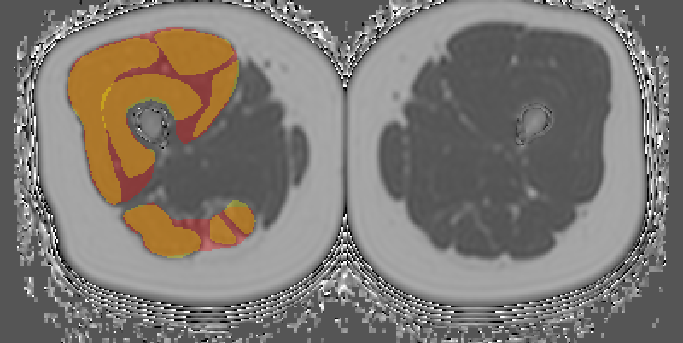
In the right thigh, axial reformations through the proximal, medial and distal third were identified. In axial PDFF maps, the entire muscle compartment as well as each compartment muscle of the hamstring (semimembranosus, semitendinosus, biceps femoris muscles) and quadriceps femoris muscle (rectus femoris, vastus lateralis, vastus medialis, vastus intermedius muscles) were manually segmented using the open-source software MITK. Mean intramuscular PDFF of single compartment muscles (PDFFm) as well as weighted averages of intra- and intermuscular PDFF of compartments (PDFFc) were extracted (Fig. 1). Intermuscular PDFF of a compartment was calculated by the formula $$PDFF_i=\frac{PDFF_c\cdot CSA_c-(PDFF_{m1}\cdot CSA_{m2}+PDFF_{m2}\cdot CSA_{m1}+...)}{CSA_c-(CSA_{m1}+CSA_{m2}+...)}$$ , with subscript ‘c’ denoting the muscle compartment and ‘m1’, ‘m2’, … corresponding compartment muscles (CSA = cross-sectional area). Four subjects with outlier PDFFi values were excluded from the statistical analysis.
Isometric Strength Measurements:
Muscle flexion and extension maximum isometric torque in Nm at the right knee joint was measured with a rotational dynamometer (Isomed 2000). Relative values in Nm³/kg were obtained by BMI-adjustment.
Results
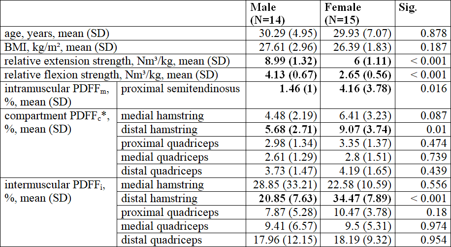
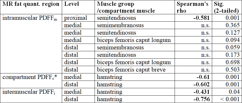
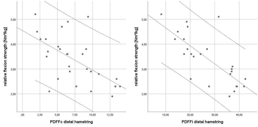
Mean relative extension and flexion strength at the right thigh was significantly higher in men than in women (Tab. 1). In the proximal level, no hamstring intermuscular PDFFi was calculated, because only the semitendinosus muscle was included. Intramuscular PDFFm of proximal semitendinosus as well as compartment PDFFc and intermuscular PDFFi of distal hamstring were significantly lower in men than in women. Relative flexion strength was significantly correlated with proximal semitendinosus PDFFm as well as medial and distal hamstring PDFFc and PDFFi (Tab. 2, Fig. 2). Close to statistical significance, relative flexion strength was correlated with distal semimembranosus PDFFm (p=0.059). A stepwise approach of a multivariate linear regression model including all intra-, intermuscular and compartment PDFF measures yielded intermuscular PDFFi of distal hamstring as the single most important factor correlated with relative flexion strength. There was no statistically significant (p<0.05) correlation between relative extension strength and any PDFF measure of the quadriceps muscle or its compartment muscles.Discussion and Conclusion
Independent of sampling in the proximal, medial or distal level of the right thigh, compartment average and intermuscular PDFF were significantly associated with flexion strength measurements in healthy subjects. Intermuscular PDFFi of distal hamstring did slightly improve the association to flexion strength beyond compartment PDFFc. These findings are particularly important for clinical application of water-fat MR, as the rather time-consuming segmentation procedure could be limited to the entire muscle compartment in a single thigh region.Acknowledgements
The present work was supported by the European Research Council (grant agreement No 637164 – iBack and 677661 ‑ ProFatMRI) and Philips Healthcare. This work reflects only the authors’ view and the EU is not responsible for any use that may be made of the information it contains.References
1. Dahlqvist JR, Vissing CR, Thomsen C, et al. Severe paraspinal muscle involvement in facioscapulohumeral muscular dystrophy. Neurology 2014;83:1178–83.
2. Kumar D, Karampinos DC, MacLeod TD, et al. Quadriceps intramuscular fat fraction rather than muscle size is associated with knee osteoarthritis. Osteoarthr Cartil 2014;22:226–34.
3. Hu HH, Kan HE. Quantitative proton MR techniques for measuring fat. NMR Biomed 2013;26:1609–29.
4. Baum T, Inhuber S, Dieckmeyer M, et al. Association of Quadriceps Muscle Fat With Isometric Strength Measurements in Healthy Males Using Chemical Shift Encoding-Based Water-Fat Magnetic Resonance Imaging. J Comput Assist Tomogr 2016;40:447–51.
5. Schlaeger S, Inhuber S, Rohrmeier A, et al. Association of paraspinal muscle water-fat MRI-based measurements with isometric strength measurements. Eur Radiol https://doi.org/10.1007/s00330-018-5631-8.
6. Karampinos DC, Yu H, Shimakawa A, et al. T₁-corrected fat quantification using chemical shift-based water/fat separation: application to skeletal muscle. Magn Reson Med 2011;66:1312–26.
Figures