1173
Resting-state fMRI at UHF: Optimizing BOLD sensitivity by using plug-and-play parallel transmission at 7T1Neurospin, CEA, Gif-sur-Yvette, France, 2Department of Cognitive Neuroscience, Faculty of Psychology and Neuroscience, Faculty of Psychology and Neuroscience, Maastricht University, Maastricht, Netherlands, 3Center for Magnetic Resonance Research, University of Minnesota, Minneapolis, MN, United States
Synopsis
The 7T Human Connectome Project (HCP) resting-state fMRI (RS-fMRI) protocol employs slice-accelerated multiband (MB) EPI with a total of 10-fold acceleration (MB 5 and in-plane 2). Such highly accelerated acquisitions provide exquisite temporal resolution at good sensitivity in most cortical regions, but quickly become very SNR starved in transmit field (B1+) deprived regions. This can be mitigated using parallel RF transmission (pTX), which however usually comes at the expense of time-consuming calibrations and pulse computations. This work shows experimentally with HCP-style RS-fMRI scans that a plug-and-play alternative for B1+ mitigation using pTX is possible with multiband Universal Pulses.
Introduction
For the 7T Human Connectome Project (HCP) resting-state fMRI (RS-fMRI) protocol, important sequence developments have been done to enable short repetition times while maintaining good spatial resolutions1,2. However, for such whole-brain studies at 7T, the transmit B1+ field heterogeneity can still lead to severe local deterioration of the signal, hence temporal SNR (tSNR) and BOLD sensitivity. Recently, application of the parallel transmission (pTX) technology3 has been proposed to solve this problem4. Specifically, the SMS-EPI sequence used in HCP was modified to enable slice-specific RF shimming. Importantly, there, RF shim weights were obtained by numerical optimization from measured subject-specific B1+ maps. In this work, by extending the Universal Pulse (UP) concept5 to SMS multi-band excitations, we propose yet another alternative solution where such calibrations are made unnecessary. In this approach, to better cope with inter-subject B1+ variability, a broader class of pTX pulses than RF shims must be considered6. In this work, we hence use a multi-band bipolar two-spoke pTX pulse design7–10 and validate our method with HCP-style RS-fMRI scans on five healthy adults.Methods
Measurements on N=5 subjects were performed on a Magnetom 7T Siemens (Siemens Healthineers, Erlangen, Germany) scanner equipped with the Nova Medical (Wilmington, MA, USA) 8TX-32RX head coil, under local SAR supervision. The RS-fMRI protocol consisted of a 15min fat-suppressed SMS-EPI acquisition with following parameters: 90 axial slices of 1.6 mm thickness (no gap), FOV=(208mm)2, matrix size 1302, in plane resolution 1.6mm, flip angle (FA) 45°, TR=1s, TE=22ms, MB=5, FOV/3 CAIPIRINHA shift, in-plane GRAPPA acceleration=2 with FLEET reference scans, partial Fourier=7/8, readout bandwidth=1832 Hz/pixel). This protocol was repeated on each subject three times on different days, to compare (i) regular single-channel transmission (sTX), (ii) sTX with dielectric (CaTiO3) padding11 and (iii) Universal Pulses pTX. B1+ maps were also acquired to retrospectively validate excitation performance by Bloch simulation (Normalized Root Mean Square (NRMS) deviation of the flip angle and nominal GRE signal). The multiband UP design consisted of slice-specific bipolar two-spoke pulses and made use of a database of 10 B1+ and B0 maps, obtained in a previous study6. For each slice, the spokes RF weights and k-space positions12 were optimized so as to minimize the FA-NRMS deviation from the target FA, averaged across the field maps database6. For tSNR calculation, the MB-EPI acquisition series were first corrected for rigid body motion using FSL McFLIRT and for distortion using Topup. Analysis of the resting-state data was carried out with nilearn and consisted in standardizing the time varying signals to unit-variance, linear detrending and band-pass filtering (0.01-0.1Hz), and regressing out the motion correction parameters and the physiological-noise-related confounds with CompCor. For all voxels, the Pearson correlation coefficient relative to a seed placed in the posterior cingulate cortex was then calculated to obtain a representation of the default mode network13 (DMN).Results
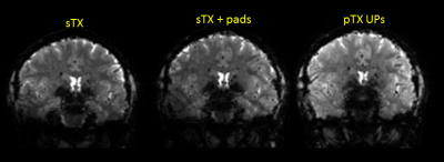
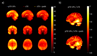
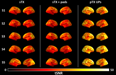
The retrospective FA simulations maps indicate that the NRMS deviation of about 25% in regular sTX acquisition is reduced to only ~10% when using universal 2-spoke pTX excitations (Table 1), an amelioration that is well exemplified by the native image comparison shown in Figure 1. In terms of signal deviation, this translates to a very moderate 4-6% using pTX-UPs, and a much larger (>14%) deviation in sTX. The dielectric pads were helpful in compensating for B1+ drops in their direct vicinity, but resulted in minimal reduction of the NRMS error due to the global nature of the NRMS metric. Temporal SNR distributions are displayed in Figures 2-3 to demonstrate a marked gain using pTX mainly for the lower brain. Interestingly also, and in agreement with the tSNR analysis, the seed-based DMN analysis indicate stronger time-correlations between the posterior cingulate cortex and the temporal cortex in the data acquired with pTX-UPs (Figure 4).Conclusions
Calibration-free pTX was successfully implemented in a HCP-style RS-fMRI protocol at 7T through the computation of slice-specific bipolar two-spoke penta-band UPs and their integration into a SMS-EPI sequence. With this work, we report for the first time universal multi-band pTX spokes pulses capable of enhancing BOLD sensitivity (up to 2-fold tSNR gains were reported) in B1+ deprived regions. This constitutes a promising outlook on whole-brain BOLD fMRI at ultra-high field.Acknowledgements
This research received funding from the European Research Council under the European Union’s Seventh Framework Program (FP7/2013-2018), ERC Grant Agreement n. 309674. B.A.P. is funded by the Netherlands Organization for Scientific Research (NWO 016.Vidi.178.052) and the National Institute of Health (R01MH111444, PI Feinberg). X.W. was supported by NIH grants U01 EB025144 and P41 EB015894 (PI K. Ugurbil).References
1. Moeller, S. et al. Multiband multislice GE-EPI at 7 tesla, with 16-fold acceleration using partial parallel imaging with application to high spatial and temporal whole-brain fMRI. Magn. Reson. Med. 63, 1144–1153 (2010).
2. Setsompop, K. et al. Blipped-controlled aliasing in parallel imaging for simultaneous multislice echo planar imaging with reduced g-factor penalty. Magn. Reson. Med. 67, 1210–1224 (2012).
3. Katscher, U., Börnert, P., Leussler, C. & van den Brink, J. S. Transmit SENSE. Magn Reson Med 49, 144–150 (2003).
4. Wu, X. et al. Human Connectome Project-style resting-state functional MRI at 7 Tesla using radiofrequency parallel transmission. NeuroImage 184, 396–408 (2019).
5. Gras, V., Vignaud, A., Amadon, A., Le Bihan, D. & Boulant, N. Universal pulses: A new concept for calibration-free parallel transmission. Magn. Reson. Med. 77, 635–643 (2017).
6. Gras, V. et al. Homogeneous non-selective and slice-selective parallel-transmit excitations at 7 Tesla with universal pulses: A validation study on two commercial RF coils. PLOS ONE 12, e0183562 (2017).
7. Saekho, S., Yip, C., Noll, D. C., Boada, F. E. & Stenger, V. A. Fast-kz three-dimensional tailored radiofrequency pulse for reduced B1 inhomogeneity. Magn. Reson. Med. 55, 719–724 (2006).
8. Setsompop, K. et al. Slice-selective RF pulses for in vivo B1+ inhomogeneity mitigation at 7 Tesla using parallel RF excitation with a 16-element coil. Magn. Reson. Med. 60, 1422–1432 (2008).
9. Tse, D. H. Y., Wiggins, C. J. & Poser, B. A. Estimating and eliminating the excitation errors in bipolar gradient composite excitations caused by radiofrequency-gradient delay: Example of bipolar spokes pulses in parallel transmission. Magn. Reson. Med. 78, 1883–1890 (2018).
10. Gras, V. et al. New method to characterize and correct with sub-µs precision gradient delays in bipolar multispoke RF pulses. Magn. Reson. Med. 78, 2194–2202 (2017).
11. Webb, A. G. Dielectric materials in magnetic resonance. Concepts Magn. Reson. Part A 38A, 148–184 (2011).
12. Gras, V. et al. In vivo demonstration of whole-brain multislice multispoke parallel transmit radiofrequency pulse design in the small and large flip angle regimes at 7 Tesla. 78, 1009–1019 (2016)
13. Vincent, J. L. et al. Coherent Spontaneous Activity Identifies a Hippocampal-Parietal Memory Network. J. Neurophysiol. 96, 3517–3531 (2006).
Figures


