1164
Gadoxetate acid disodium-enhanced MRI: Multiple arterial phases using differential sub-sampling with cartesian ordering (DISCO) may achieve more optimal late arterial phases than the single arterial phase imaging1West China Hospital, Sichuan University, Chengdu, China, 2GE Healthcare China, MR Research, Beijing, China
Synopsis
Differential sub-sampling with cartesian ordering (DISCO) is a high spatial-temporal resolution MR technique that combines multiple features: a dual echo SPGR echo acquisition for DIXON water-fat separation, a pseudo-random variable density k-spacesegmentation, parallel imaging and a view sharing reconstruction. Gadoxetate acid disodium is a specific hepatobiliary contrast agent, however, the high frequency happening of transient severer motion can impair arterial phase (AP) images. In this study, we prospectively determine multiple AP imaging using DISCO can improve the capturing rate of well-timed late AP with less motion artifact in in comparison to single AP imaging using LAVA-Flex.
Synopsis
Differential sub-sampling with cartesian ordering (DISCO) is a high spatial-temporal resolution MR technique that combines multiple features: a dual echo SPGR echo acquisition for DIXON water-fat separation, a pseudo-random variable density k-spacesegmentation, parallel imaging and a view sharing reconstruction. Gadoxetate acid disodium is a specific hepatobiliary contrast agent, however, the high frequency happening of transient severer motion can impair arterial phase (AP) images. In this study, we prospectively determine multiple AP imaging using DISCO can improve the capturing rate of well-timed late AP with less motion artifact in in comparison to single AP imaging using LAVA-Flex.Purpose
To prospectively determine whether the use of a multiple arterial phase imaging technique (DISCO) improve the capturing rate of late arterial phase with less motion artifact than single arterial phase obtained with gadoxetate acid disodiumMaterials and methods
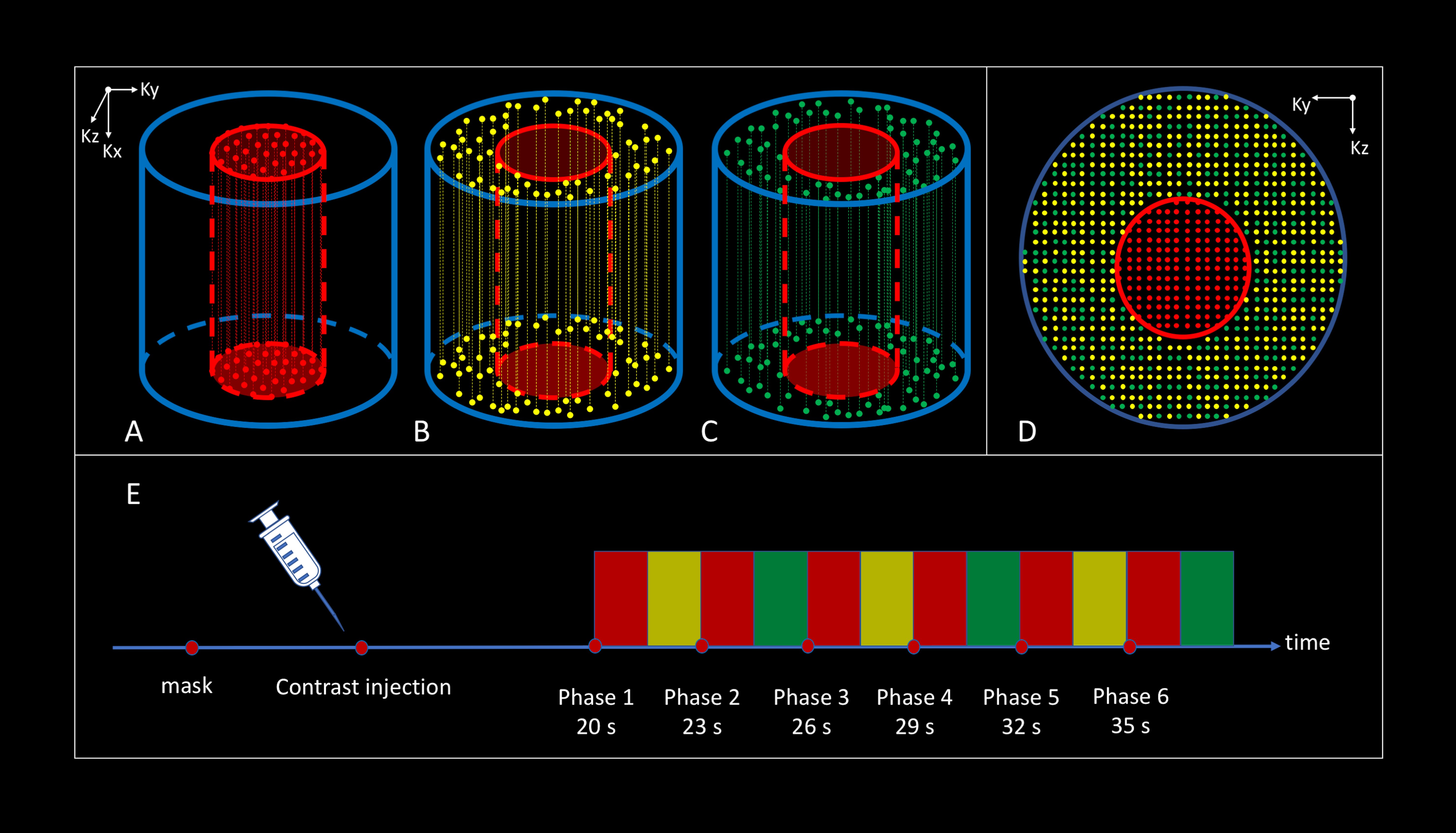
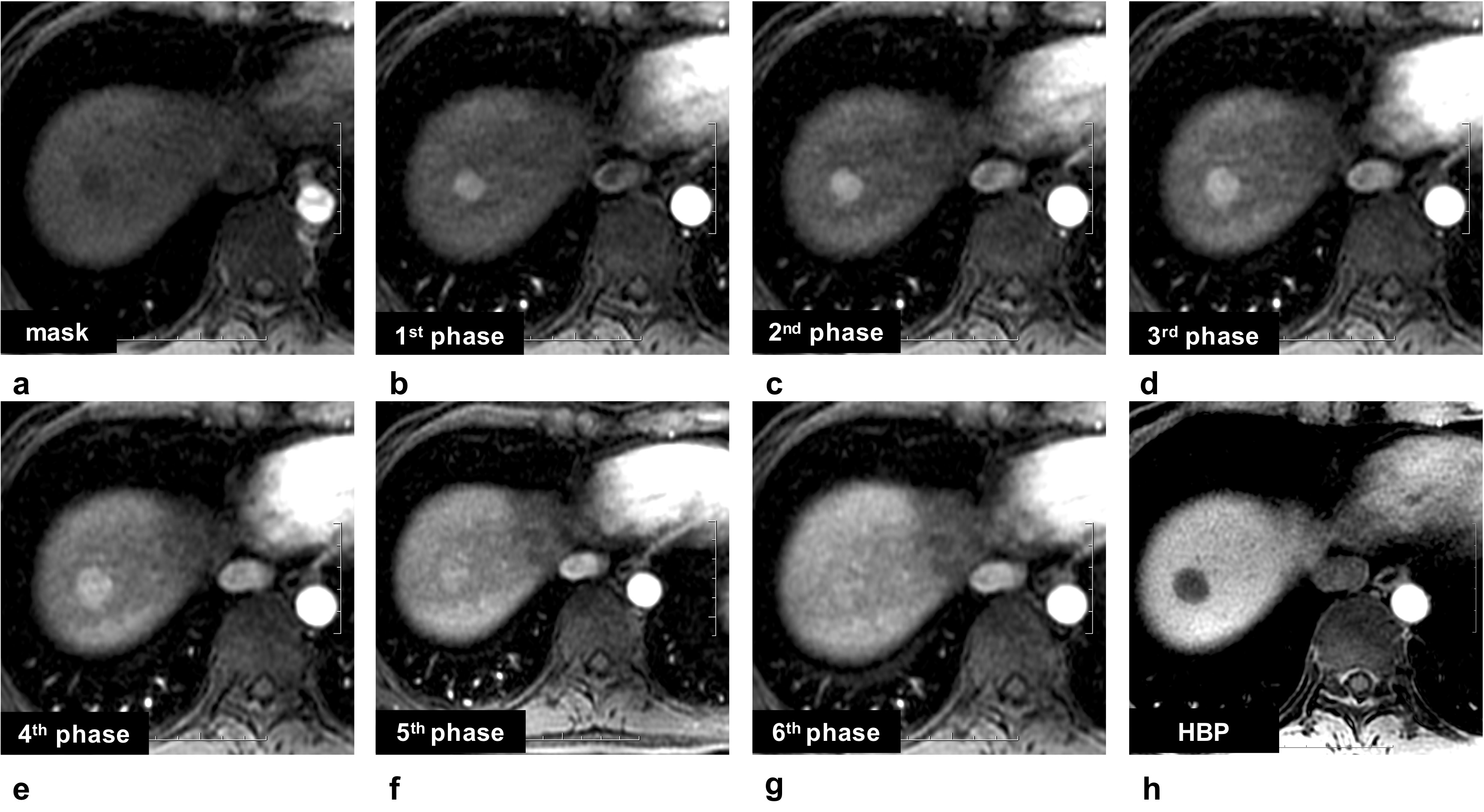
In this institutional review board-approved study, prospectively acquired data of 132 patients who underwent either single (n=67) or multiple arterial phase (n=65) gadoxetate acid disodium-enhanced MR imaging from 08/2017 to 07/2018 were analyzed. MR was carried out by using a 3.0 T MR system (Discovery MR 750w, GE Healthcare, Milwaukee, USA). A sixteen-channel phased-array torsor coil (GE Medical System) was used for all examinations. The multiple arterial phase imaging were obtained using differential sub-sampling with cartesian ordering (DISCO) by dividing the k-space data into 3 regions:1 center region and 2 outer regions. The center regions are acquired every other acquisition while the outer groups are acquired less frequently and were shared with the nearest center regions for the final image reconstruction (Figure 1).For the subcohort A patients, multiple arterial phase images were acquired in an 18 s long breath hold with a 20 s delay after the injection of gadoxetate acid disodium (Primovist, Bayer Pharma AG, Berlin, Germany) with the injection rate of 1 ml/s (Figure 1). These data were reconstructed into sixth sequential phases with a temporal resolution of 3 s. Since the center k-space region were sampled every time and outer region were subsampled, the center k-space region images were reconstructed at 20 s, 23s, 26 s, 29 s, 32 s, and 35 s (Figure 2). For the subcohort B patients, the single arterial phase images were acquired with a 20 s delay after the contrast injection. Two readers independently assessed the arterial phase timing and the degree of motion artifact on a five-point scale. The kappatest was used to determine the agreement between the two readers,c2 or fisherexact test were used for the categorical variables and Student ttest or Mann-Whitney Utest were used for the comparison of the motion artifacts between the single arterial phase and the multiple arterial phase imaging.Results
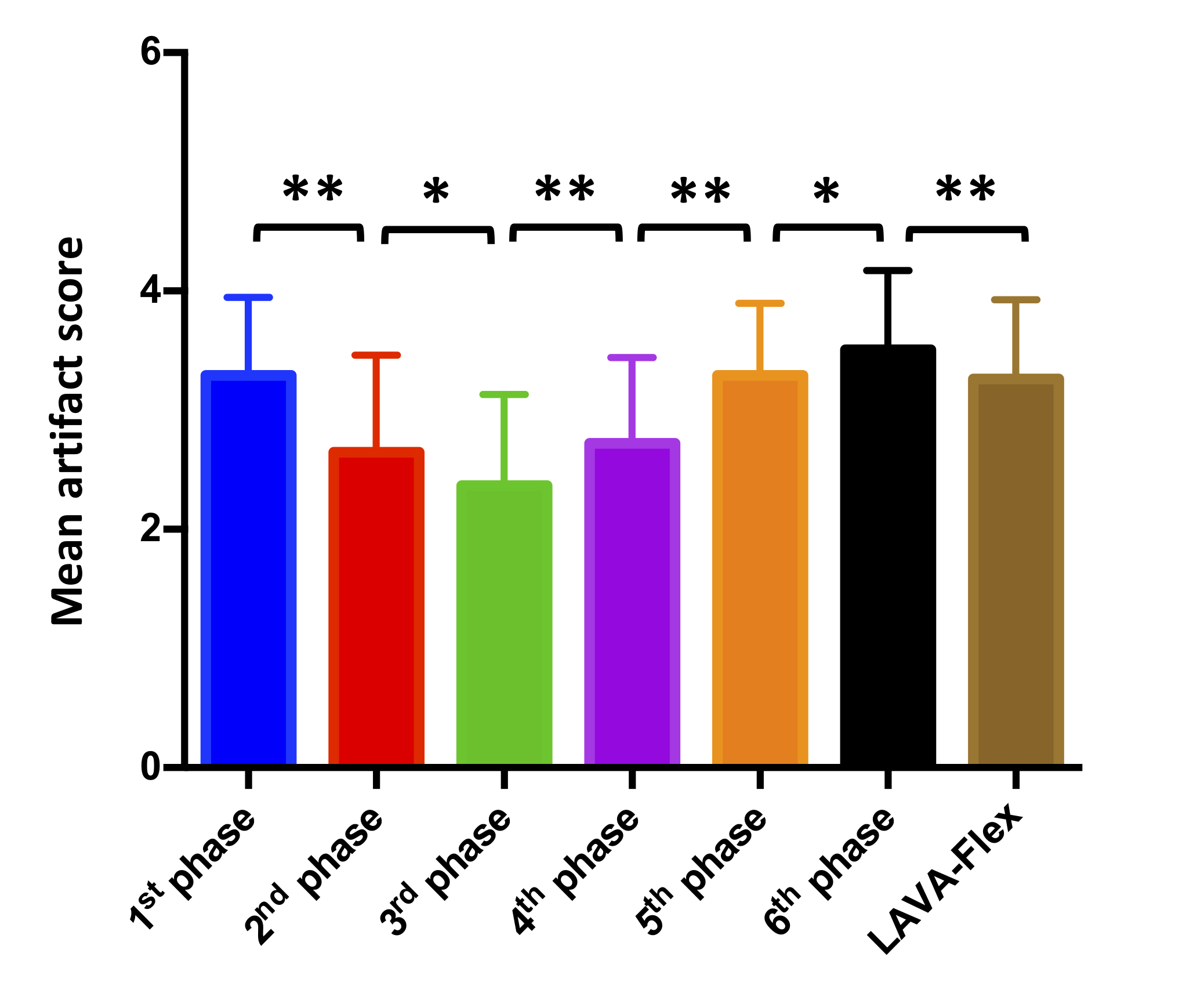
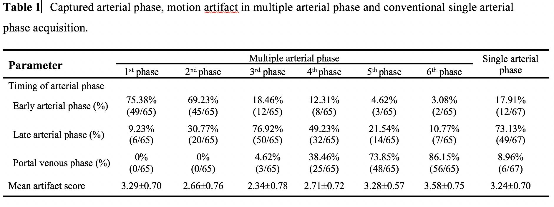
Good to perfect inter-observer agreement was obtained for the arterial phase timing and degree of motion artifact (all kappavalue > 0.70). Optimal timing of arterial phase was observed in 95.4% (62/65) of multiple arterial phase acquisitions compared with 73.1% (49/67) of single arterial phase acquisitions (c2=12.209, p<0.001) (Table 1). Motion artifact score of the late arterial phase images measured using single arterial phase acquisition (3.22±0.68) was significant higher than the multiple arterial phase (2.42±0.74) group (t=5.921,p<0.001). For the multiple arterial phase acquisition, motion artifact score of the 2nd (2.66±0.76), 3rd(2.34±0.78) and 4th (2.71±0.72) phases were also significant reduced compared with 1st(3.29±0.70), 5th (3.28±0.57) and 6th (3.58±0.75) phases (all p<0.001), and statistical significance was also found between 2ndand 3rd (p=0.01), 3rdand 4th(p=0.003) phases(Figure 3).Conclusion and Discussion:
The results of this study demonstrated that the use of multiple arterial phase acquisition with DISCO captured well-timed hepatic late arterial phase images more frequently than single arterial phase imaging with LAVA-Flex. Furthermore, less motion artifacts were observed of the late arterial phase images using multiple arterial phase imaging compared with the single arterial phase imaging. Thus, multiple hepatic arterial phase acquisition may result in more optimal late arterial phase and provide high image quality with less motion artifacts.Acknowledgements
No acknowledgement found.References
1. Duncan JK, Ma N, Vreugdenburg TD, et al. Gadoxetic acid-enhanced MRI for the characterization of hepatocellular carcinoma: A systematic review and meta-analysis. J Magn Reson Imaging. 2017;45(1):281-90.
2. McInnes MD, Hibbert RM, Inacio JR, Schieda N. Focal Nodular Hyperplasia and Hepatocellular Adenoma: Accuracy of Gadoxetic Acid-enhanced MR Imaging-A Systematic Review. Radiology. 2015;277(3):927.
3. Jiang HY, Chen J, Xia CC, et al. Noninvasive imaging of hepatocellular carcinoma: From diagnosis to prognosis. World J Gastroenterol. 2018;24(22):2348-62.
4. Joo I, Lee JM, Lee DH, et al. Noninvasive diagnosis of hepatocellular carcinoma on gadoxetic acid-enhanced MRI: can hypointensity on the hepatobiliary phase be used as an alternative to washout? Eur Radiol. 2015;25(10):2859-68.
5. Motosugi U, Bannas P, Bookwalter CA, et al. An Investigation of Transient Severe Motion Related to Gadoxetic Acid-enhanced MR Imaging. Radiology. 2016;279(1):93-102.
6. Yoon JH, Lee JM, Yu MH, et al. Evaluation of Transient Motion During Gadoxetic Acid-Enhanced Multiphasic Liver Magnetic Resonance Imaging Using Free-Breathing Golden-Angle Radial Sparse Parallel Magnetic Resonance Imaging. Invest Radiol. 2018;53(1):52-61
Figures