0933
A novel method combining Dixon water-fat separation and BLADE imaging1Siemens Shenzhen Magnetic Resonance Ltd., Shenzhen, China, 2Siemens Medical Solutions USA, Boston, MA, United States
Synopsis
Fat suppression and motion artifacts are still challenging in clinical MRI for certain body regions. In this work a novel acquisition method combining Dixon’s method and BLADE (or PROPELLER, periodically rotated overlapping parallel lines with enhanced reconstruction) is proposed, to address fat suppression and motion artifacts simultaneously. We show that the proposed technique acquires data efficiently while avoiding the potential misalignment between echoes as well as the phase inconsistency introduced by different readout directions.
Introduction
MRI often suffers from both B0 inhomogeneity related artifact and motion artifact in critical body regions. For example, B0 inhomogeneity in the neck results in incomplete fat suppression and even water suppression, while swallowing and arterial pulsation may introduce motion artifacts. Dixon’s method and BLADE (or PROPELLER) have been used widely to address these problems separately. Dixon’s method is a model-based water-fat separation technique which is more robust to B0 inhomogeneity. However, they are usually implemented in Cartesian k-space acquisitions, which is prone to motion artifact. On the other hand, BLADE sequence is able to reduce motion artifact, so various strategies combining Dixon with BLADE have been proposed [1-4]. Turboprop IDEAL [1] method samples several echoes around each spine echo using EPI readout for Dixon water-fat separation, which is very efficient but requires additional correction to remove phase errors arising from the EPI readout. Another class of methods using fly-back gradients [2, 3] to avoid the phase errors but may still suffer from motion, as for each blade the echoes required by Dixon model are acquired in different shots. A method recently proposed by Schär et al. addressed the phase error and potential motion by acquiring two echoes with same readout direction in one shot [4].
In this work, we propose a novel method combining BLADE and Dixon. In this new approach, two blade groups and two echoes for each group are acquired per shot. Water and fat blades are produced after blade-by-blade Dixon reconstruction. Then BLADE reconstruction is performed on the water and fat blades separately to generate final water and fat images.
Methods
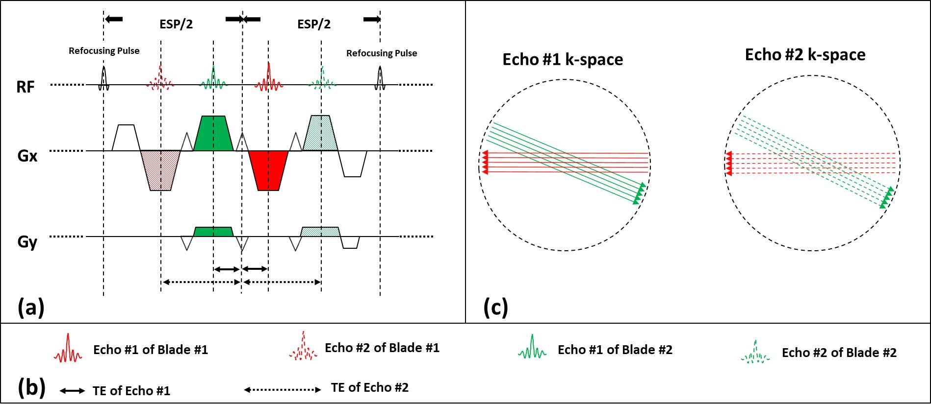
Contrast to conventional BLADE, in this method multiple blades are acquired in one shot. One spin echo period is shown in Figure 1a. The echoes marked with solid-red and dashed-red lines are collected at the same k-space location, with same readout direction but at difference echo time. Similarly, the echoes marked with solid-green and dashed-green lines are collected at another k-space location to form a second group of blades. Such acquisition is repeated throughout the whole echo train with different phase encoding steps, producing all k-space lines for each blade. The k-space locations of these blades are shown in Figure 1c.
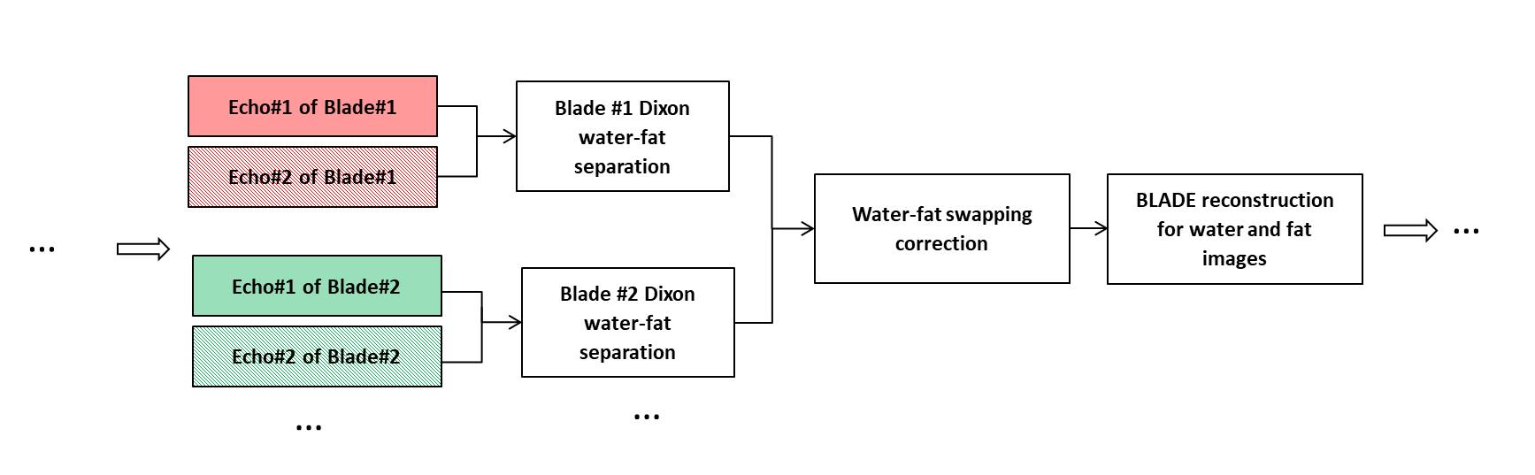
The reconstruction pipeline is shown in Figure 2. Dual-echo Dixon reconstruction [5] is performed on the two echoes of each blade, generating water and fat signals for each blade. Then the water and fat blades are sent to standard BLADE reconstruction to generate full k-space water and fat images, respectively.
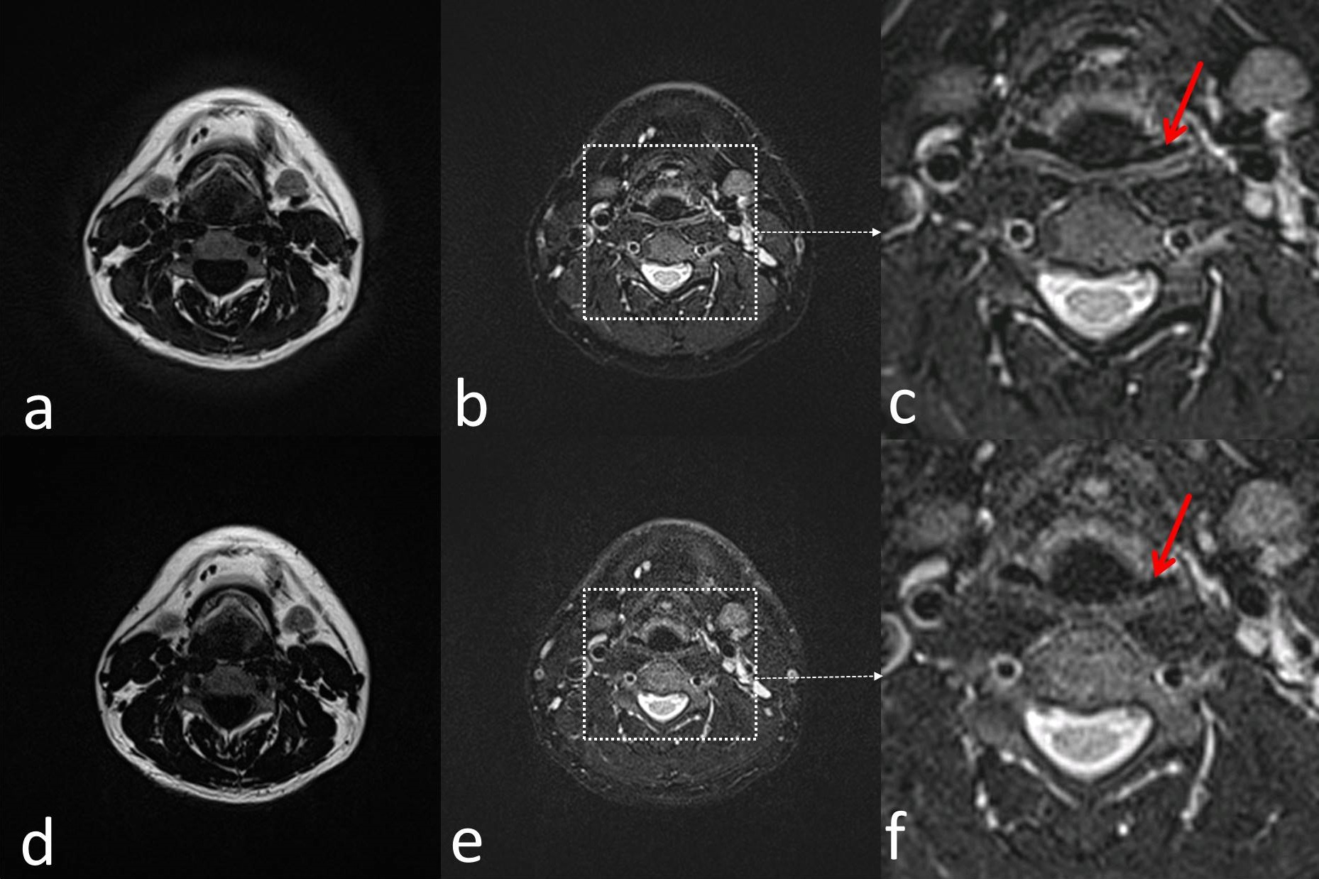
The proposed method was tested in the neck region of a healthy volunteer at a Siemens 1.5T MAGNETOM Aera scanner (Siemens Healthineers, Erlangen, Germany) with a T2-weighted protocol. The following parameters were used: 0.9×0.9×3 mm3 resolution, 40 slices, and bandwidth = 780 Hz/Pixel, TE/TR = 84ms/6300ms, measurement time = 2’58’’. For comparison, product TSE Dixon was acquired with matching resolution and number of slices. The different parameters are: bandwidth = 820 Hz/Pixel, TE/TR = 78ms/4500ms, measurement time = 3’04’’.
Results
Figure 3 show one out of 40 slices acquired with the proposed Dixon BLADE sequence (a-c) and product TSE Dixon sequence (d-f). Comparison of the fat and water images to the TSE Dixon sequence shows that correct water-fat separation was achieved by the proposed Dixon BLADE sequence. Due to pulsation and other involuntary motion, e.g., swallowing, some artifact and blurring are visible in the region around vertebra on the TSE Dixon water image. In the proposed sequence, BLADE trajectory suppressed these artifacts and resulted in improved image quality (b, c).Discussion and Conclusion
In this work, a novel method combing Dixon and BLADE is demonstrated. The advantages of this acquisition strategy are three fold: First, the potential misalignment between different echoes is avoided as the two echoes are acquired in the same TR; second, in addition, the two echoes are acquired with the same readout gradient polarity, so that water-fat separation can be performed without phase correction; third, the high efficiency of data acquisition, i.e., almost all the time between refocusing pulses are used for sampling data except for the readout pre-phasing and re-phasing gradients.Acknowledgements
The authors thank Dr. Dominik Nickel and Alto Stemmer for the valuable discussions regarding this work.References
[1] Huo D, Li Z, Aboussouan E, Karis JP, Pipe JG. Turboprop IDEAL: a motion-resistant fat-water separation technique. Magn Reson Med 2009;61:188–195.
[2] Weng D, Pan Y, Zhong X, Zhuo Y. Water–fat separation with parallel imaging based on BLADE. Magn Reson Imaging 2013;31:656–663.
[3] He Q, Weng D, Zhou X, Ni C. Regularized iterative reconstruction for undersampled BLADE and its applications in three-point Dixon water-fat separation. Magn Reson Med 2011;65:1314–1325.
[4] Schär M, Eggers H, Zwart NR, Chang Y, Bakhru A, Pipe JG. Dixon water‐fat separation in PROPELLER MRI acquired with two interleaved echoes. Magnetic resonance in medicine. 2016 Feb;75(2):718-28.
[5] Eggers H, Brendel B, Duijndam A, Herigault G., Dual-echo Dixon imaging with flexible choice of echo times. Magnetic resonance in medicine. 2011 Jan;65(1):96-107.
Figures