0925
Real-time shimming in the cervical spinal cord using an 8-channel AC/DC array and a capacitive respiratory sensor1NeuroPoly Lab, Institute of Biomedical Engineering, Polytechnique Montréal, Montreal, QC, Canada, 2Electrical Engineering and Computer Science, Massachusetts Institute of Technology, Cambridge, MA, United States, 3Athinoula A. Martinos Center for Biomedical Imaging, Massachusetts General Hospital, Charlestown, MA, United States, 4Harvard Medical School, Boston, MA, United States, 5Harvard-MIT Division of Health Sciences and Technology, Cambridge, MA, United States, 6Functional Neuroimaging Unit, CRIUGM, University of Montreal, Montreal, QC, Canada
Synopsis
Spinal cord imaging remains challenging in large part due to the temporal variations in B0 caused by respiration. This study introduces a simple circuit design for a capacitive respiratory sensor and demonstrates the utility of this device in the context of real-time shimming the neck using an 8-channel "AC/DC" (integrated RF receive + multi-coil shim) array. Using this approach, motion artifacts in a multi-echo T2*-weighted acquisition are shown to be considerably reduced.
Introduction
Spinal cord MRI is hampered by static and respiration-induced resonance offsets (RIRO)1, both of which cause artifacts in echo-planar (EPI) and gradient-echo (GRE) based acquisitions. Recent investigations have demonstrated that by using respiratory bellows, along with a series of B0 field measurements, RIRO effects in the spinal cord can to a large extent be compensated either prospectively2 by real-time shimming and/or retrospectively3 during image reconstruction. While these studies are encouraging, the respiratory trace on which the corrections are based has been shown to be an imperfect surrogate for fluctuations in B04, e.g. whether the bellows are more or less sensitive to abdominal or chest forms of breathing depends on how they are fit to a given subject. NMR field probes5 are an attractive alternative, but they are generally expensive and require extensive additions to hardware. In this study, we introduce a simple design for an alternative respiratory sensor based on capacitive coupling and apply it, in combination with an integrated 8-channel "AC/DC" coil6, to real-time shimming in the cervical spinal cord.Methods
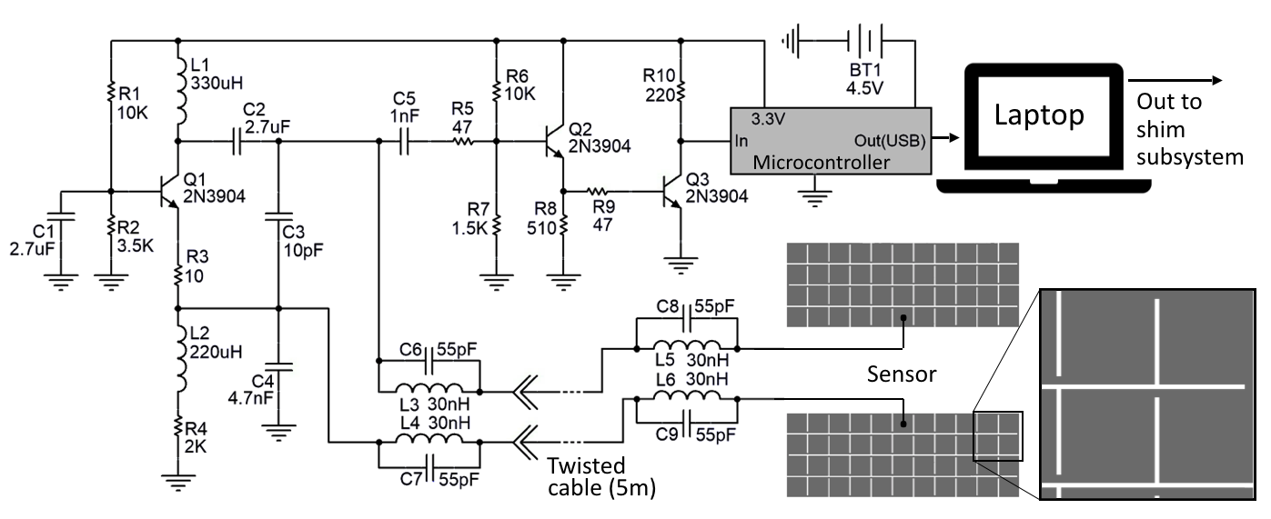
The respiratory monitor (Figure 1) was based on a Colpitts oscillator and a capacitive sensor which is part of the capacitive voltage divider of the resonant tank circuit7,8. The sensor consists of two rectangular copper plates which were segmented into strips to minimize gradient-induced eddy currents using the LF Frequency Domain Solver of CST Studio Suite. To minimize RF interference during excitation, parallel resonant circuits, tuned to 123.2 MHz, were inserted along the twisted pair connecting the sensor to the oscillator.
The sensor is positioned atop the patient bed and beneath a subject's lungs. Under loading (human subject), a capacitance of approximately 250 pF was measured, with a maximum range of 14 pF between inspired and expired conditions. As the dielectric properties of the thorax vary during respiration, the capacitance of the sensor changes which, in turn, modulates the operating frequency of the oscillator. A Teensy microcontroller (version 3.5) measures the frequency and writes the value to USB, which is then read by the computer controlling the shim subsystem.
Experiments
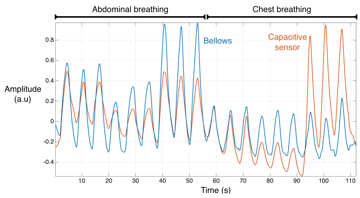
An initial comparison was performed outside of the scanner between the capacitive sensor and traditional bellows. With the subject lying supine atop the capacitive sensor and the bellows strapped around their abdomen, the subject was instructed to perform 3 cycles of measured breathing with 3 different intensities: 3 normal breaths, followed by 3 shallow breaths, followed by 3 deep breaths. This sequence was performed first while breathing abdominally and, once again, while breathing predominantly from the chest. Respiratory signals from the two detectors were recorded simultaneously.
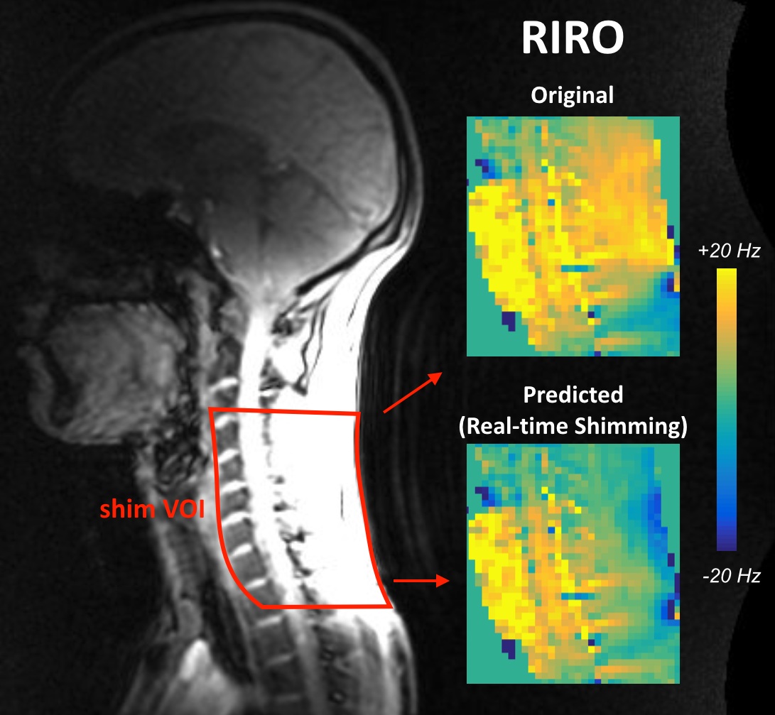
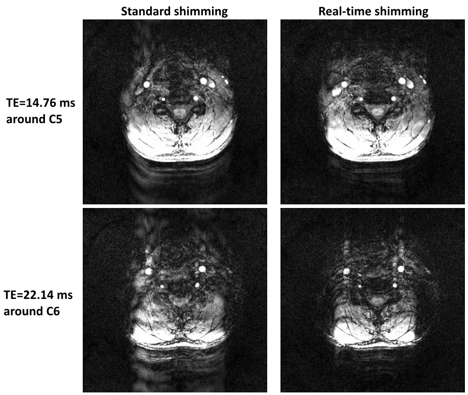
As proof of concept, real-time shimming using an 8-channel AC/DC coil6,9 (i.e. each RF Rx element doubles as a local shim coil) was applied to an axial 3D multi-echo T2*-weighted acquisition centered at the C5/C6 vertebral disk using the following parameters: TE = [4.06, 8.57, 14.76, 22.14 ms], TR = 27 ms, 20-degree flip angle, readout along the left-right axis, and voxel size (FOV/matrix size) = [190/288 mm, 190/288 mm, 104/30 mm]. To calibrate the real-time updates to shim current2, sagittal GRE-field maps were first acquired under inspired and expired breath-holds while concurrently recording the respiratory trace, yielding an estimate of how B0 changes along with respiration (Figure 2). A shim volume of interest (shim VOI) was automatically derived from the GRE magnitude using the Spinal Cord Toolbox10: A cylinder of radius 40 mm centered along the centerline of the spinal cord was extended between vertebral levels C2 to T2 and, because the in-plane phase encoding was performed along the AP axis, voxels within the back but posterior to the cylindrical volume were also included. Shim currents for the 8 channels (constrained to 2.5 A absolute current per channel) were optimized to reduce RIRO across the shim VOI.
During the T2*-weighted scans, the respiratory signal was sampled at 20 Hz and shims were updated every 100 ms based on the two preceding respiratory samples. The subject was instructed to breathe normally during the scan which, upon completion, was repeated again after turning the AC/DC coil off and shimming anew with the static spherical harmonic shims of the scanner (up to 2nd order).
Results and Discussion
Experimental results are shown in Figure 3 and Figure 4. We have demonstrated the utility of a capacitive respiratory sensor to inform real-time shim adjustments. Future work will investigate how well such a sensor might generalize for different body types (loading conditions) and, critically, the extent of its correlation to the B0 field variations targeted by real-time shimming.Acknowledgements
Funded by the Canada Research Chair in Quantitative Magnetic Resonance Imaging [950-230815], the Canadian Institute of Health Research [CIHR FDN-143263], the Canada Foundation for Innovation [32454, 34824], the Fonds de Recherche du Québec - Santé [28826], the Fonds de Recherche du Québec - Nature et Technologies [2015-PR-182754, B2X doctoral scholarship], the Natural Sciences and Engineering Research Council of Canada [435897-2013], the Canada First Research Excellence Fund (IVADO and TransMedTech) and the Quebec BioImaging Network [5886].References
1. Verma, T. & Cohen-Adad, J. Effect of respiration on the B0 field in the human spinal cord at 3T. Magn Reson Med72,1629–36 (2014).2. Topfer, R., Foias, A., Stikov, N. & Cohen-Adad, J. Real-time correction of respiration-induced distortions in the human spinal cord using a 24-channel shim array. Magn Reson Med80,935–946 (2018).
3. Vannesjo, S. J., Clare, S., Kasper, L., Tracey, I. & Miller, K. L. Trace-based correction of breathing-induced field fluctuations in T2 * -weighted imaging of the spinal cord. bioRxiv(2018). doi:10.1101/406371
4. Vannesjo, S. J., Miller, K. L., Clare, S. & Tracey, I. Spatiotemporal characterization of breathing-induced B0 field fluctuations in the cervical spinal cord at 7T. Neuroimage167,191-202 (2018).
5. De Zanche, N., Barmet, C., Nordmeyer-Massner, J. a. & Pruessmann, K. P. NMR Probes for measuring magnetic fields and field dynamics in MR systems. Magn Reson Med60,176–186 (2008).
6. Topfer, R. et al.Integrated ΔB0/Rx coil array for improved spinal cord imaging at 3T. in Proceedings of the 26th Annual Meeting of ISMRM, Paris, France, 2018. Abstract 8930.
7. Aponte Luis, J, Roa Romero, L.M., Gómez-Galán, J.A., Hernández, D. N., Estudillo-Valderrama, M. A., Barbarov-Rostán, G, & Rubia-Marcos, C. Design and Implementation of a Smart Sensor for Respiratory Rate Monitoring. Sensors14,3019–3032 (2014).
8. Oum, J. H., Koo, H. & Hong, S. Non-contact Heartbeat Sensor using LC oscillator circuit. in 30th Annual International IEEE EMBS Conference4455–4458 (2008).
9. Stockmann, J. P. et al.A 32-channel combined RF and B0 shim array for 3T brain imaging. Magn Reson Med75,441–451 (2016).
10. De Leener, B. et al.SCT: Spinal Cord Toolbox, an open-source software for processing spinal cord MRI data. Neuroimage145,24–43 (2016).
Figures