0698
Free-breathing Volumetric Body Imaging for Combined Qualitative and Quantitative Tissue Assessment using MR MultitaskingZixin Deng1, Anthony G. Christodoulou1, Nan Wang1, Wensha Yang2, Lixia Wang3, Yi Lao2, Fei Han4, Xiaoming Bi4, Bin Sun5, Stephen J Pandol6, Richard Tuli2, Debiao Li1, and Zhaoyang Fan1
1Biomedical Imaging Research Institute, Cedars-Sinai Medical Center, Los Angeles, CA, United States, 2Department of Radiation Oncology, Cedars-Sinai Medical Center, Los Angeles, CA, United States, 3Radiologic Department, Chaoyang Hospital, Beijing, China, 4Siemens Healthineers, Los Angeles, CA, United States, 5Department of Radiology, Fujian Medical University Union Hospital, Fuzhou, China, 6Department of Medicine, Cedars-Sinai Medical Center, Los Angeles, CA, United States
Synopsis
MR has broad clinical applications in body imaging with excellent soft-tissue contrast and qualitative and emerging quantitative assessments, however,
Introduction
MR has broad clinical applications in body imaging. Excellent soft-tissue contrast with conventional tissue property-weighted imaging and emerging quantitative assessment with advanced mapping methods have made MR advantageous over other imaging modalities. However, respiratory motion represents a critical constraint on the capability and quality of body MR. MR multitasking is a recently developed method capable of resolving overlapping dynamics such as respiratory motion, T1 relaxation, and T2 relaxation [1]. In this work, we propose a free-breathing body imaging technique based on MR Multitasking to provide respiratory phase-resolved multi-task images including T1-, T2-weighted images and their corresponding maps.Methods
The proposed multitasking technique features innovations in both acquisition and reconstruction. K-space is sampled continuously using a prototype stack-of-stars gradient echo sequence with golden angle ordering in-plane and Gaussian-density randomized ordering in the partition direction, interleaved with training data (0° in-plane, central partition) every 10th readout. A saturation recovery (SR) preparation and length-variable T2 preparations or an inversion preparation are combined to generate T1-weighted and T2-weighted signals during magnetization evolution (Figure 1). Multi-task images (respiratory-resolved 3D T1-, T2-weighted and T1-, T2-maps) are generated based on a low-rank tensor image reconstruction framework. The proposed method was tested in an ISMRM/NIST phantom, 6 healthy subjects, and 5 cancer patients (2 pancreas, 3 liver) at 3.0T. For the in vitro study, standard inversion recovery spin echo imaging at various TIs and spin echo imaging at various TEs were used for reference T1 and T2 mapping, respectively. For healthy in vivo studies, T1 and T2 values from the multitasking method was compared to published literature values [2-4] in various abdominal organs. Specific imaging parameters for all multitasking scans were: TE/TR = 3.1/6.0ms, spatial resolution = 1.7x1.7x6.0mm3, FOV = 275x275x312mm3, whole abdominal coverage with 52 partitions in the axial direction, flip angle = 5° (following SR preparation)/10° (following T2 or inversion preparation), water-excitation for fat suppression. Total imaging time was approximately 10 minutes.Results
In vitro studies: excellent correlation was observed between T1 reference and T1 multitasking (R2=0.998) and between T2 reference and T2 multitasking (R2=0.933) in the ISMRM/NIST MRI system phantom (Figure 2). In vivo studies: the proposed technique provided spatially co-registered multi-task 3D image sets in all subjects (Figure 3). Figure 4 shows a liver cancer patient and a pancreatic cancer patient example. In healthy subjects, T1 values from the multitasking method showed close agreement with the literature values in the liver, spleen, pancreas, skeletal muscle, but were slightly higher in the kidney. T2 values from the multitasking method were comparable to T2 values from literature (Table 1). Overall, higher T1 and T2 values were observed in tumorous regions (pancreatic and liver) compared to healthy organs (Table 1).Discussion & Conclusion
This study demonstrated the feasibility of a free-breathing volumetric, quantitative body imaging technique based on MR Multitasking. Respiratory phase-resolved multi-task images including T1-, T2-weighted images and corresponding maps can be acquired simultaneously within a single 10-minute scan. Good image quality and comparable T1, T2 measurements to reference scans or published literature were observed both in phantom and human studies. The proposed multitasking method has the advantage of providing flexible image contrast, qualitative and quantitative respiratory-resolved information for diagnosis and staging of cancer, longitudinal tumor response monitoring, and MR-guided radiotherapy planning.Acknowledgements
No acknowledgement found.References
[1] Christodoulou AG, Shaw JL, Nguyen C, Yang Q, Xie Y, Wang N, Li D. “Magnetic resonance multitasking for motion-resolved quantitative cardiovascular imaging,” Nature Biomed Eng 2018. [2] de Bazelaire CMJ, Duhamel GD, Rofsky NM, Alsop DC. MR imaging relaxation times of abdominal and pelvic tissuesm easured in vivo at 3.0 T: preliminary results. Radiology 2004;230(3):652–659. [3] Chen Y, Lee GR, Aandal G, et al. Rapid volumetric T1 mapping of the abdomen using three-dimensional through-time spiral GRAPPA. Magn Reson Med 2015 May 18. [4] Stanisz GJ, Odrobina EE, Pun J, et al. T1, T2 relaxation and magnetization transfer in tissue at 3T. Magn Reson Med 2005;54(3):507–512.Figures
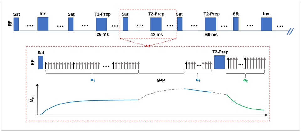
Figure
1. Pulse sequence design to obtain T1 and T2 measurements. Saturation recovery
(SR) pulse is interleaved with inversion recovery and various T2-preparation
(T2prep) modules ranging from 26ms, 42ms, and 66ms. The cycle is then repeated.
A Flip angle of 5 degrees (𝜶1) was used during the
SR phase and a flip angle of 10 degrees (𝜶2) was used during the
T2-prepared phase.
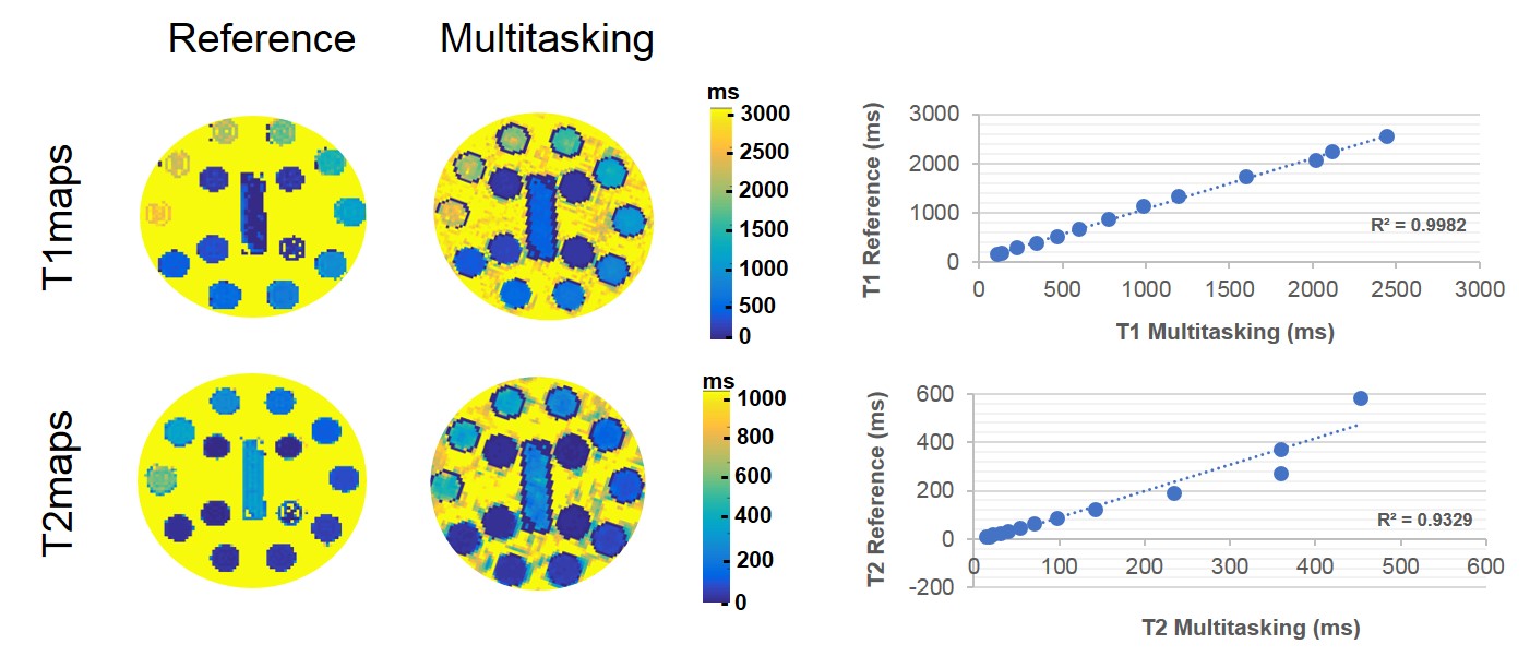
Figure
2. The ISMRM/NIST MRI system phantom validation. Correlation plots comparing T1
and T2 values between the proposed multitasking technique and the conventional
inversion-recovery spin echo and spin echo methods. Note that, T2 values above
200ms are not typically seen in abdominal organs.
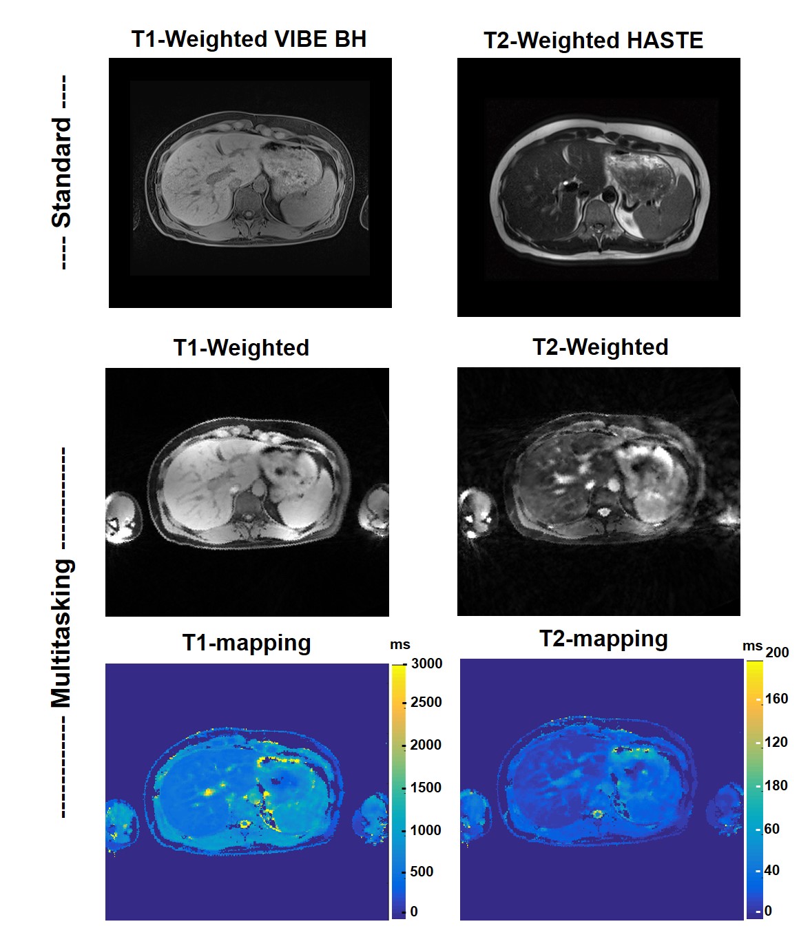
Figure
3. Healthy subject. Top: standard T1-weighted breath-hold VIBE and T2-weighted
HASTE images. Bottom: 3D respiratory resolved T1-, T2-weighted and T1-, T2-maps
reconstructed using the proposed multitasking method in a healthy subject.
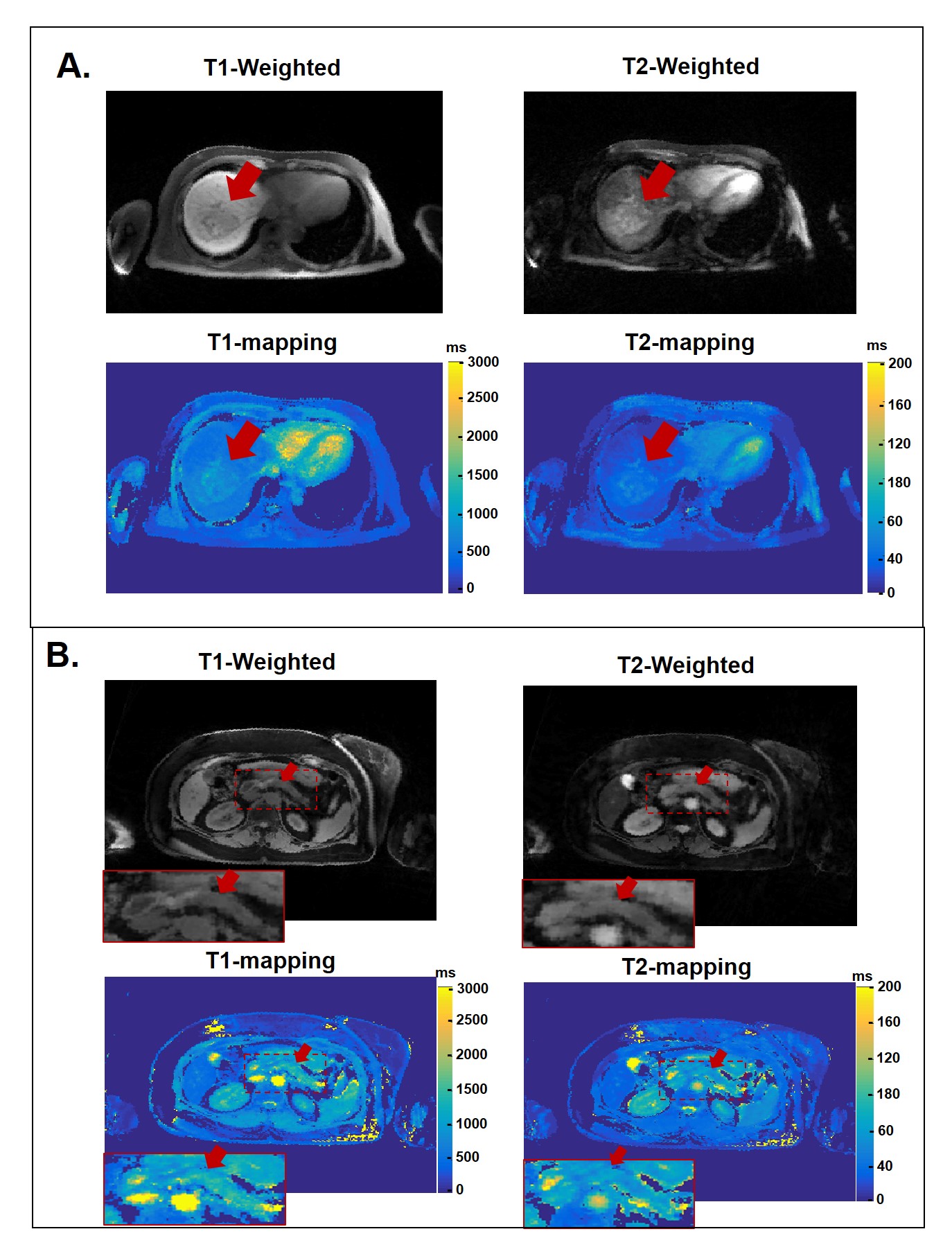
Figure 4. 3D respiratory resolved T1-, T2-weighted and T1-, T2-maps reconstructed using the proposed multitasking method in a liver cancer patient (A) and pancreas cancer patient (B).
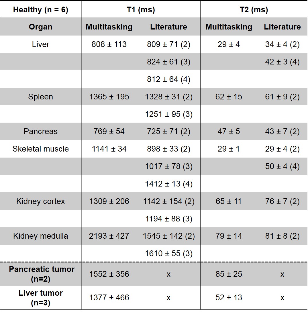
Table
1. Average T1 and T2 values of abdominal organs acquired at 3.0T. Comparison
between proposed multitasking technique and literature values.