0554
Learning-Based Jointly-Optimal Design of the Diffusion Encoding Scheme and Orientation Estimation Method for Diffusion MRI1University of Southern California, Los Angeles, CA, United States, 2Massachusetts General Hospital, Boston, MA, United States, 3Harvard Medical School, Boston, MA, United States
Synopsis
Diffusion MRI is powerful but limited by long scan times. When optimizing diffusion MRI, most previous methods have either optimized the encoding scheme (i.e., q-space samples) or have optimized the parameter estimation method. In this work, we propose and evaluate a novel approach that jointly optimizes both the encoding scheme and the estimation scheme. This is enabled by combining linear estimation theory with machine learning techniques. Our results show the strong potential of our new approach. Perhaps surprisingly and in contrast to conventional wisdom, we observe that a two-shell sampling scheme appears to be preferred for orientation estimation.
Introduction
Diffusion MRI is slow, and optimizing the diffusion encoding scheme (i.e., the q-space sampling locations) is an important task that has been studied for many years1-3. In addition, extracting a maximal amount of relevant information from a given set of q-space samples is also an important and widely-studied problem4-12. Usually, these two problems are considered independently from one another – the sampling scheme is optimized independently of diffusion parameter estimation, and vice versa. In this work, we consider a novel approach that jointly optimizes both the q-space sampling scheme and the parameter estimation scheme. Our approach specifically builds off of the previous ERFO method11-12 for parameter estimation, which combined machine learning methods with linear estimation theory to design optimized parameter estimation methods for arbitrary q-space sampling schemes. Our new approach, which we call Joint ERFO (J-ERFO) uses the same cost function and methods as ERFO, but additionally optimizes the q-space sampling locations.Theory and Methods
For simplicity, we describe J-ERFO for orientation distribution function (ODF) estimation, although note that the general approach can also be applied to other parameters. The ERFO approach11-12 assumes that ODFs will be estimated linearly from $$$M$$$ q-space samples, with $$\hat{O}(\mathbf{u})= \sum_{m=1}^M a_m (E(\mathbf{q}_m )+n_m),$$where $$$\hat{O}(\mathbf{u})$$$ is the estimated ODF, $$$E(\mathbf{q}_m)$$$ is the true diffusion signal at q-space location $$$\mathbf{q}_m$$$, $$$n_m$$$ represents noise, and the $$$a_m$$$ coefficients define the linear estimation method.
ERFO specifically determines the coefficients $$$a_m$$$ using $$$P$$$ sets of paired training data $$$(\hat{O}_p(\mathbf{u}),E_p(\mathbf{q}_m))$$$ within a machine learning framework, by optimizing $$\arg \min_{a_m} \sum_{p=1} ^P |\hat{O}_p(\mathbf{u}) - \sum_{m=1}^M a_m (E_p(\mathbf{q}_m)+n_m)|^2.$$ It has been previously demonstrated that, unlike most conventional machine learning methods, the estimation schemes learned by ERFO have strong theoretical characterizations and can generalize well to new settings that they weren’t trained for. These capabilities are related to a recently-proposed theoretical framework for understanding linear diffusion MRI methods13.
The proposed J-ERFO approach extends ERFO by also optimizing over the q-space samples: $$\arg \min_{(a_m,\mathbf{q}_m)} \sum_{p=1}^P |\hat{O}_p(\mathbf{u}) - \sum_{m=1}^M a_m (E_p(\mathbf{q}_m)+n_m)|^2.$$ While ERFO leads to a simple convex optimization problem, J-ERFO is associated with a complicated non-convex optimization problem. We find a local minimum to this problem using the variable projection approach14 and use multiple initializations to ensure that we find a relatively good local minimum. J-ERFO was trained to identify 90 q-space samples that were optimal for ODF estimation when the SNR of the unweighted (b=0 s/mm$$$^2$$$) image was 20. Training was performed for an effective diffusion time of 17.5ms ($$$\Delta=21.8$$$ ms, $$$\delta=12.9$$$ ms) based on synthetic tensor data with physiologically-realistic parameters.
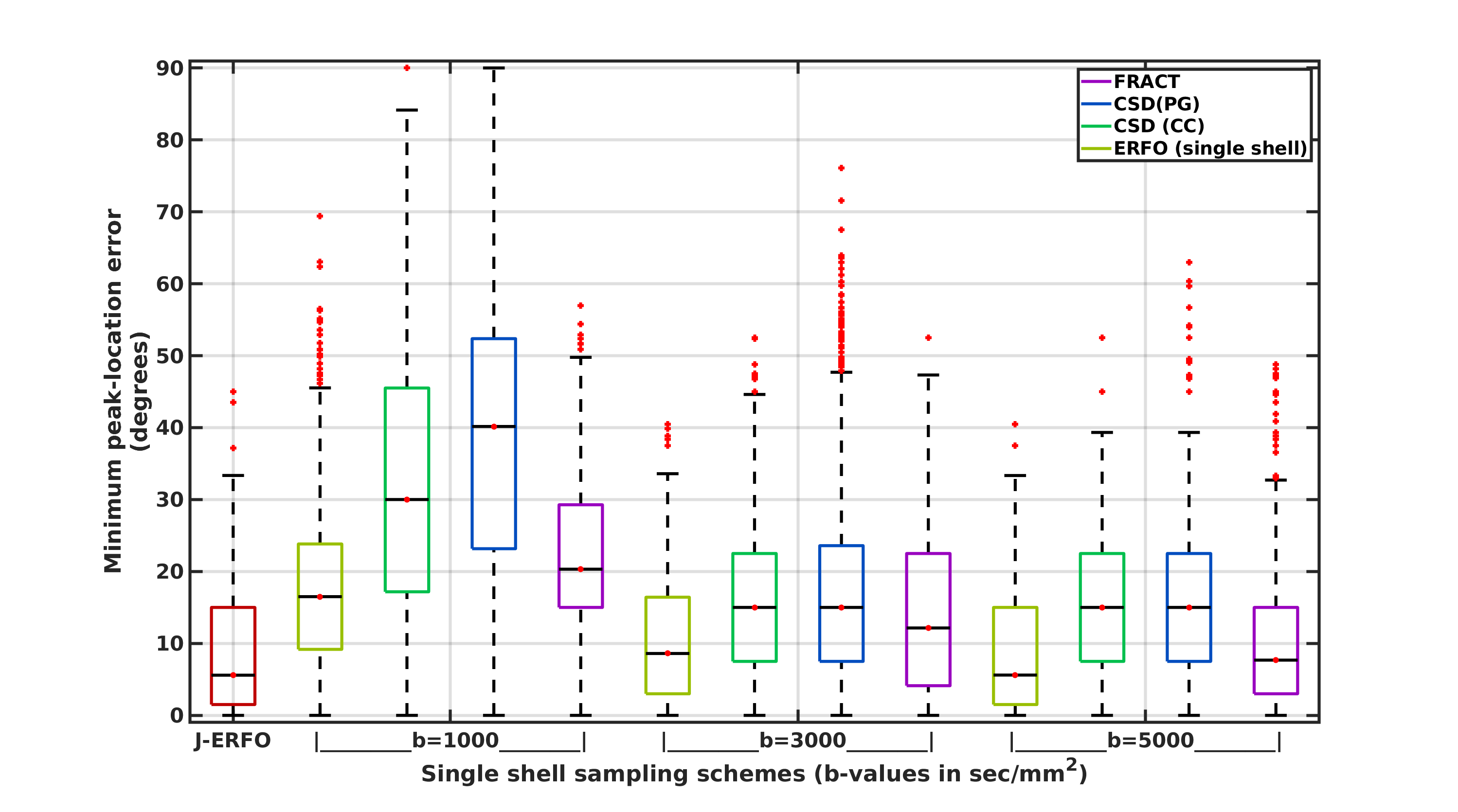
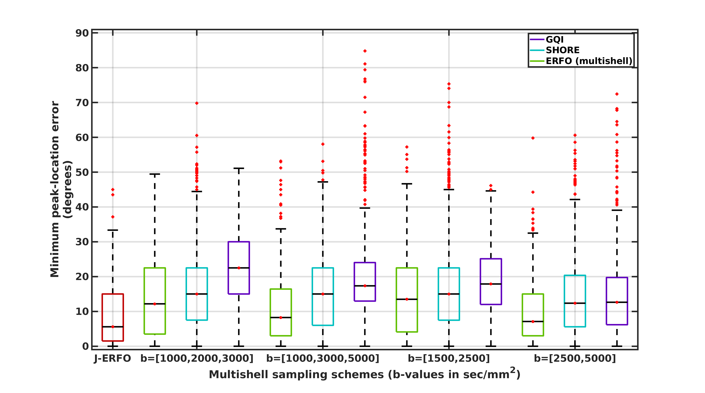
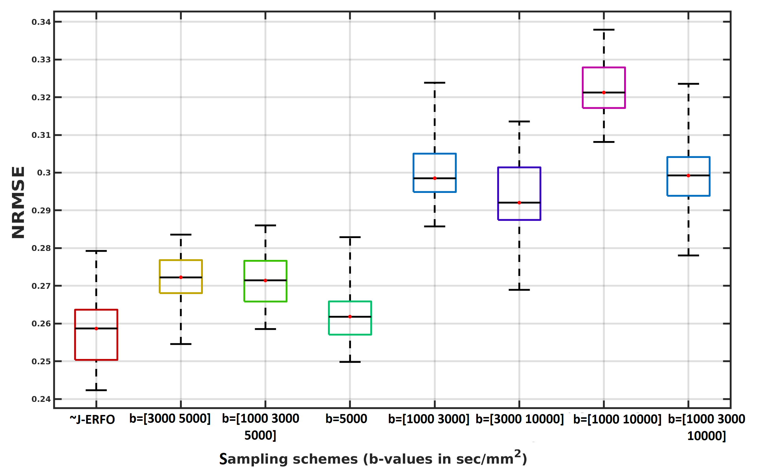
J-ERFO was tested in simulation using 2500 pairs of noisy (SNR 20) 5$$$\mu$$$m crossing cylinders15 with varying diffusivities and angles of separation. J-ERFO was compared against single shell (FRACT9, constrained spherical deconvolution (CSD)8, ERFO11-12) and multi-shell (3D-SHORE6, GQI7, ERFO11-12) ODF estimation methods. J-ERFO was also tested using real in vivo multi-shell human brain data16 (diffusion time=17.5ms and 512 images with b-values=[1000,3000,5000,10000] s/mm$$$^2$$$). An approximate J-ERFO protocol (~J-ERFO) was obtained by keeping 90 images from the dataset that best matched the J-ERFO protocol.
Results
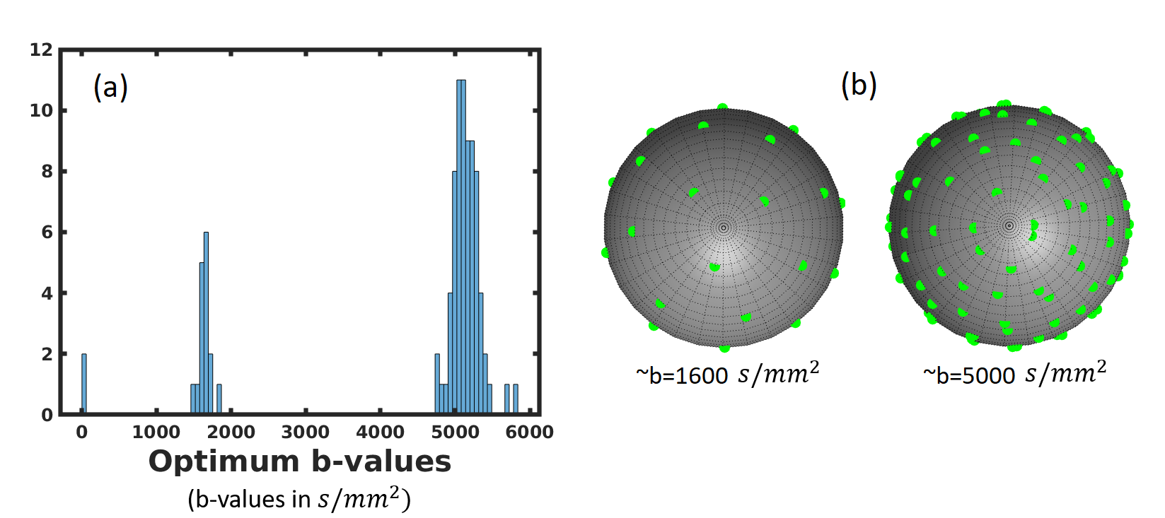
The optimal q-space sampling scheme obtained by J-ERFO is shown in Fig. 1. We observe that this approximates a two-shell scheme, with one shell at b=1600s/mm$$$^2$$$ and another shell at b=5000s/mm$$$^2$$$.
Figures 2 and 3 show comparisons against other sampling schemes and parameter estimation methods. J-ERFO consistently has the smallest error in estimating the ODF peak locations. ERFO is a close second, especially when the sampling scheme is similar to J-ERFO, which is expected behavior.
Figure 4 shows real data results from 90 q-space samples, using the full q-space data as a gold standard. ~J-ERFO has the lowest normalized root-mean-squared error (NRMSE).
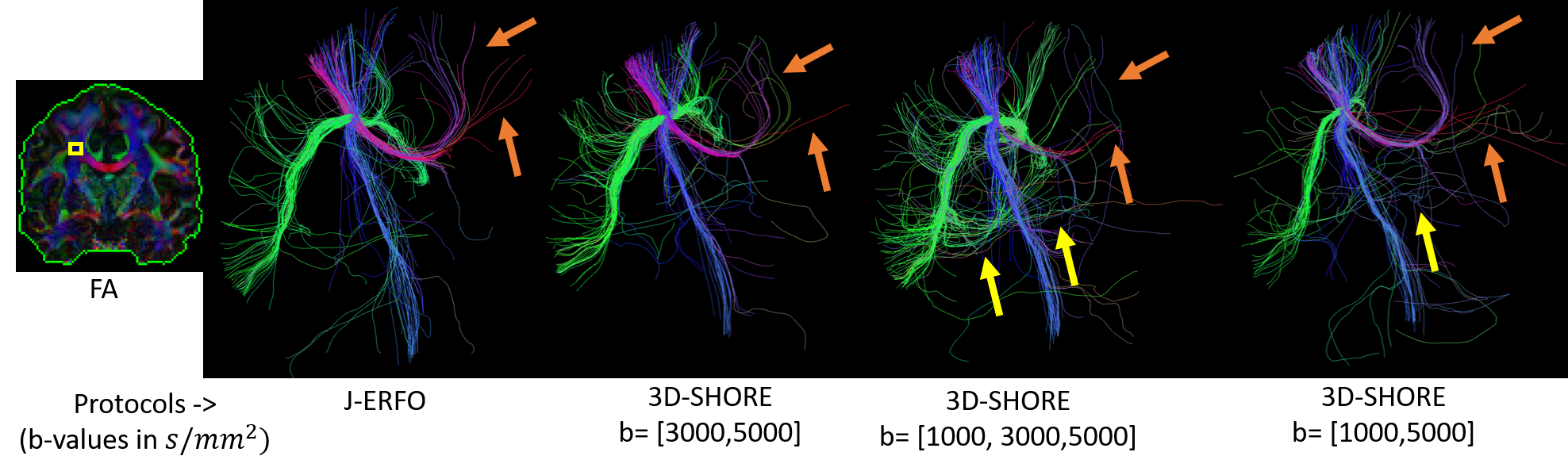
Figure 5 illustrates the tracking results of ~J-ERFO and 3D-SHORE applied to multiple multishell protocols. Only ~J-ERFO reconstructs the corpus callosum fanning (orange arrows) and shows fewer false positives than 3D-SHORE (yellow arrows).
Discussion and Conclusion
We have presented and evaluated J-ERFO, a novel learning-based method for jointly designing q-space sampling and parameter estimation methods. Our numerical simulations and in vivo evaluations all consistently suggest that J-ERFO produces better ODF estimation performance than existing methods. Interestingly, the optimized q-space sampling scheme identified by J-ERFO is distinct from many of the q-space sampling schemes described in previous literature. However, it should also be observed that the optimized b-values appearing in the J-ERFO sampling scheme can vary substantially with the assumed SNR, suggesting that the q-space sampling scheme should be tailored based on the SNR characteristics of the data.Acknowledgements
This work was supported in part by NSF CAREER award CCF-1350563 and NIH grants R01-MH116173, R01-NS074980, R01-NS089212, and R21-EB022951.
Some of the computation for the work described in this abstract was supported by the University of Southern California’s Center for High-Performance Computing (hpc.usc.edu).
References
[1] D. K. Jones, M. A. Horseld, A. Simmons, Optimal strategies for measuring diffusion in anisotropic systems by magnetic resonance imaging, Magn. Reson. Med. 42 (1999) 515-525.
[2] E. Caruyer, C. Lenglet, G. Sapiro, R. Deriche, Design of multishell sampling schemes with uniform coverage in diffusion MRI, Magn. Reson. Med. 69 (2013) 1534-1540.
[3] D. C. Alexander, A general framework for experiment design in diffusion mri and its application in measuring direct tissue-microstructure features, Magnetic Resonance in Medicine 60 (2) (2008) 439-448.
[4] V. J. Wedeen, P. Hagmann, W.-Y. I. Tseng, T. G. Reese, R. M. Weisskoff, Mapping complex tissue architecture with diffusion spectrum magnetic resonance imaging, Magn. Reson. Med. 54 (2005) 1377-1386.
[5] D. S. Tuch, Q-ball imaging, Magn. Reson. Med. 52 (2004) 1358-1372.
[6] E. Ozarslan, C. Koay, T. M. Shepherd, S. J. Blackband, P. J. Basser, Simple harmonic oscillator based reconstruction and estimation for three-dimensional q-space MRI, in: Proc. Int. Soc. Magn. Reson. Med., 2009, p. 1396.
[7] F.-C. Yeh, V. J.Wedeen, W.-Y. I. Tseng, Generalized q-sampling imaging, IEEE Trans. Med. Imag. 29 (2010) 1626-1635
[8] J.-D. Tournier, F. Calamante, D. G. Gadian, A. Connelly, Direct estimation of the fiber orientation density function from diffusion-weighted MRI data using spherical deconvolution, NeuroImage 23 (2004) 1176-1185.
[9] J. P. Haldar, R. M. Leahy, Linear transforms for Fourier data on the sphere: Application to high angular resolution diffusion MRI of the brain, NeuroImage 71 (2013) 233-247.
[10] H. Zhang, T. Schneider, C. A. Wheeler-Kingshott, D. C. Alexander, NODDI: Practical in vivo neurite orientation dispersion and density imaging of the human brain, NeuroImage 61 (2012) 1000-1016.
[11] D. Varadarajan and J. P. Haldar, Towards optimal linear estimation of orientation distribution functions with arbitrarily sampled diffusion MRI data, ISBI (2018), pp. 743-746.
[12] D. Varadarajan and J. P. Haldar, ERFO: Improved ODF estimation by combining machine learning with linear estimation theory, ISMRM (2018).
[13] D. Varadarajan and J. P. Haldar, A theoretical signal processing framework for linear diffusion MRI: Implications for parameter estimation and experiment design, Neuroimage 161 (2017) 206-218.
[14] G. Golub, V. Pereyra, Separable nonlinear least squares: the variable projection method and its applications, Inverse Probl. 19 (2003) R1-R26.
[15] L. Raguin, D. Hernando, D. Karampinos, L. Ciobanu, B. Sutton, Z. Liang, J. Georgiadis, Quantitative analysis of q-space mri data, ISMRM (2006).
[16] Q.Fan, T. Witzel, A. Nummenmaa, K. R. A. Van Dijk, J. D. Van Horn, M. K. Drews, L. H. Somerville, M. A. Sheridan, R. M. Santillana, J. Snyder, T. Hedden, E. E. Shaw, M. O. Hollinshead, V. Renvall, R. Zanzonico, B. Keil, S. Cauley, J. R. Polimeni, D. Tisdall, R. L. Buckner, V. J. Wedeen, L. L. Wald, A. W. Toga, B. R. Rosen, MGH–USC Human Connectome Project datasets with ultra-high b-value diffusion MRI, NeuroImage, 124 Part B (2016) 1108-1114.
Figures