0449
Geometric Coil Mixing (GCM) to Dampen Confounding Signals in MRI Reconstruction1Department of Radiology, A. A. Martinos Center for Biomedical Imaging, Massachusetts General Hospital, Charlestown, MA, United States, 2Harvard Medical School, Boston, MA, United States, 3Department of Physics and Astronomy, Heidelberg University, Heidelberg, Germany, 4Siemens Healthcare GmbH, Erlangen, Germany, 5Siemens Shenzhen Magnetic Resonance Ltd, Shenzhen, China, 6Fetal-Neonatal Neuroimaging & Developmental Science Center, Boston Children's Hospital, Boston, MA, United States, 7Division of Neuroradiology, Department of Radiology, Massachusetts General Hospital, Boston, MA, United States, 8Harvard-MIT Health Sciences and Technology, Massachusetts Institute of Technology, Cambridge, MA, United States
Synopsis
We introduce an artifact reduction technique that exploits the spatial locality afforded by multi-channel receiver array coils. Specifically, we create an optimal coil mixing with the purpose of dampening confounding signals prior to parallel imaging (PI) reconstruction. We demonstrate the mitigation of artifacts caused by PI model inaccuracies for Wave-CAIPI imaging in neurological and MSK applications. In addition, we illustrate the potential of this technique for minimizing the effects of non-rigid maternal and fetal motion during fetal brain imaging. This computationally efficient approach should allow for direct application of model based reconstruction/motion correction methods in difficult imaging scenarios.
Introduction
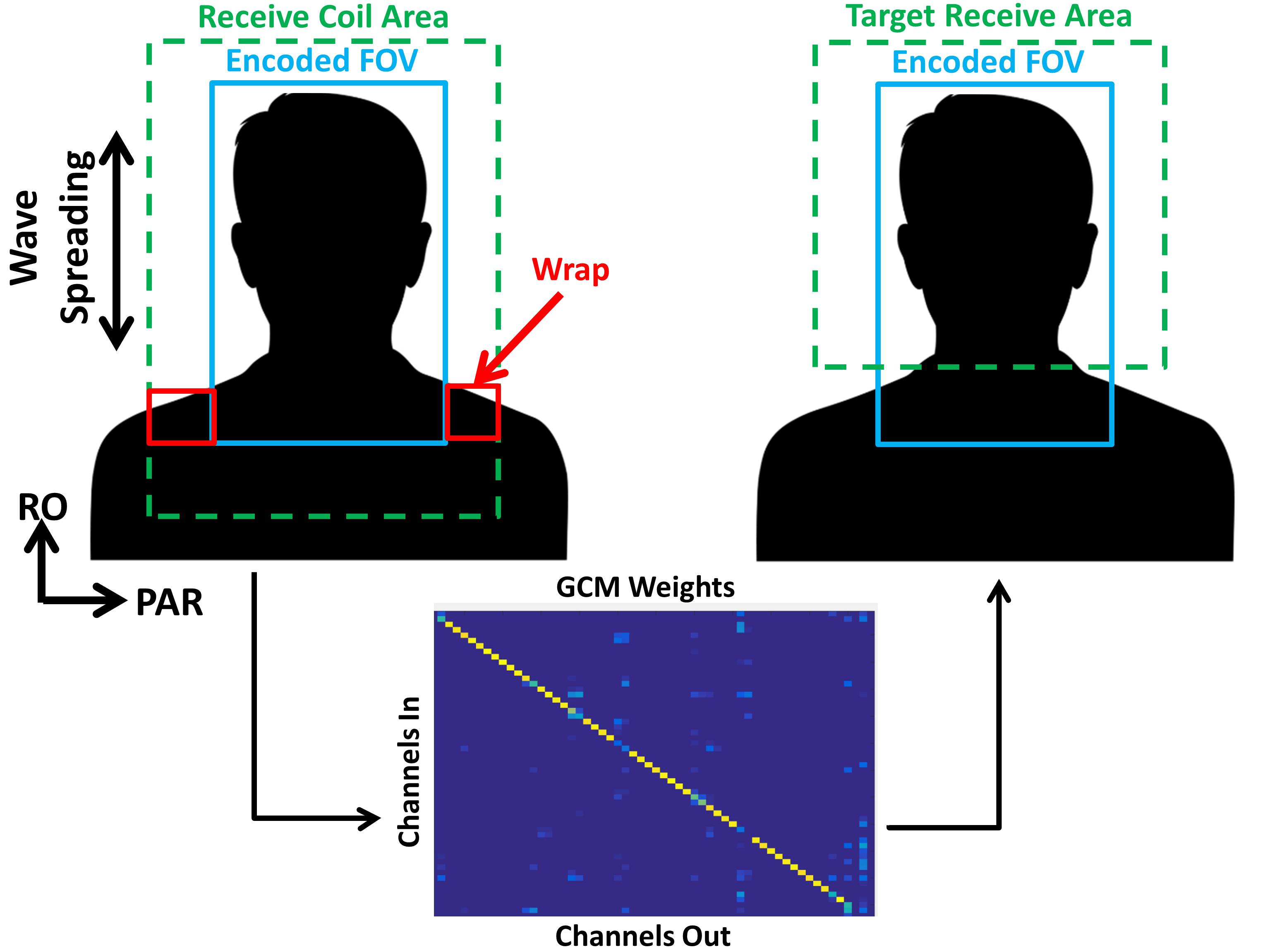
The advancement of parallel imaging (PI)1–4 and other model based reconstruction methods5,6 for highly accelerated imaging has been greatly aided by advances in multi-channel receiver array coils7,8. Many of these advanced reconstruction approaches have a substantial computational burden which has lead researchers to explore software and hardware based model reduction techniques9–12. These techniques attempted to remove redundancy in the acquired data to reduce the computational cost of the reconstruction. A geometric coil compression technique12 improved the compression efficiency of Cartesian data by exploiting spatial locality properties inherent to array coils. These locality properties had also served as the foundation for one of the initial PI techniques (PILS)3. Alternatively, in this work we leverage the locality properties of the coils to remove undesirable signal sources prior to applying model based reconstruction or motion correction. This is accomplished by solving for the best linear coil mixing (Geometric Coil Mixing, GCM) matrix that attempts to restrict the coil sensitivities to a region of interest. The estimation process can be efficiently performed on the auto-calibration data. The mixing is then applied to the accelerated data to mitigate artifacts that are caused by model inaccuracies or data corruption from sources outside of the region of interest. Fig. 1 illustrates the input/output relationship used to determine the GCM for a neurological imaging application.
Methods
The proposed GCM was implemented within an online reconstruction of the Wave-CAIPI13 acquisition scheme (available through Siemens C2P sharing program for T1w, T2w SPACE, FLAIR, and T1w MPRAGE sequences). In compliance with IRB requirements, two healthy adult volunteers and a pregnant patient were scanned on 3T MRI Scanners (MAGNETOM Prisma, Siemens Healthcare, Erlangen, Germany). T1w MPRAGE Wave-CAIPI data were acquired at 1mm iso resolution, R=3×4, 256mm3 FOV, [TR, TI, TE] = [2500, 1100, 3.5]ms, and BW=195Hz/px using a 64ch head/neck coil. T2w SPACE Wave-CAIPI data were acquired at 0.625×0.625×0.6mm3 resolution, R=2×2, FOV 160×160×124mm3, [TR, TE] = [900, 26]ms, BW = 405Hz/px using an 18ch knee coil. The GCM strategy was also tested offline in MATLAB (Mathworks, MA, USA) using fetal HASTE data acquired at 1.3×1.3×3mm3 resolution, 330×330×108mm3 FOV, [TR, TE] = [1600, 117]ms, BW = 700Hz/px using a 36ch body/spine coil.Results
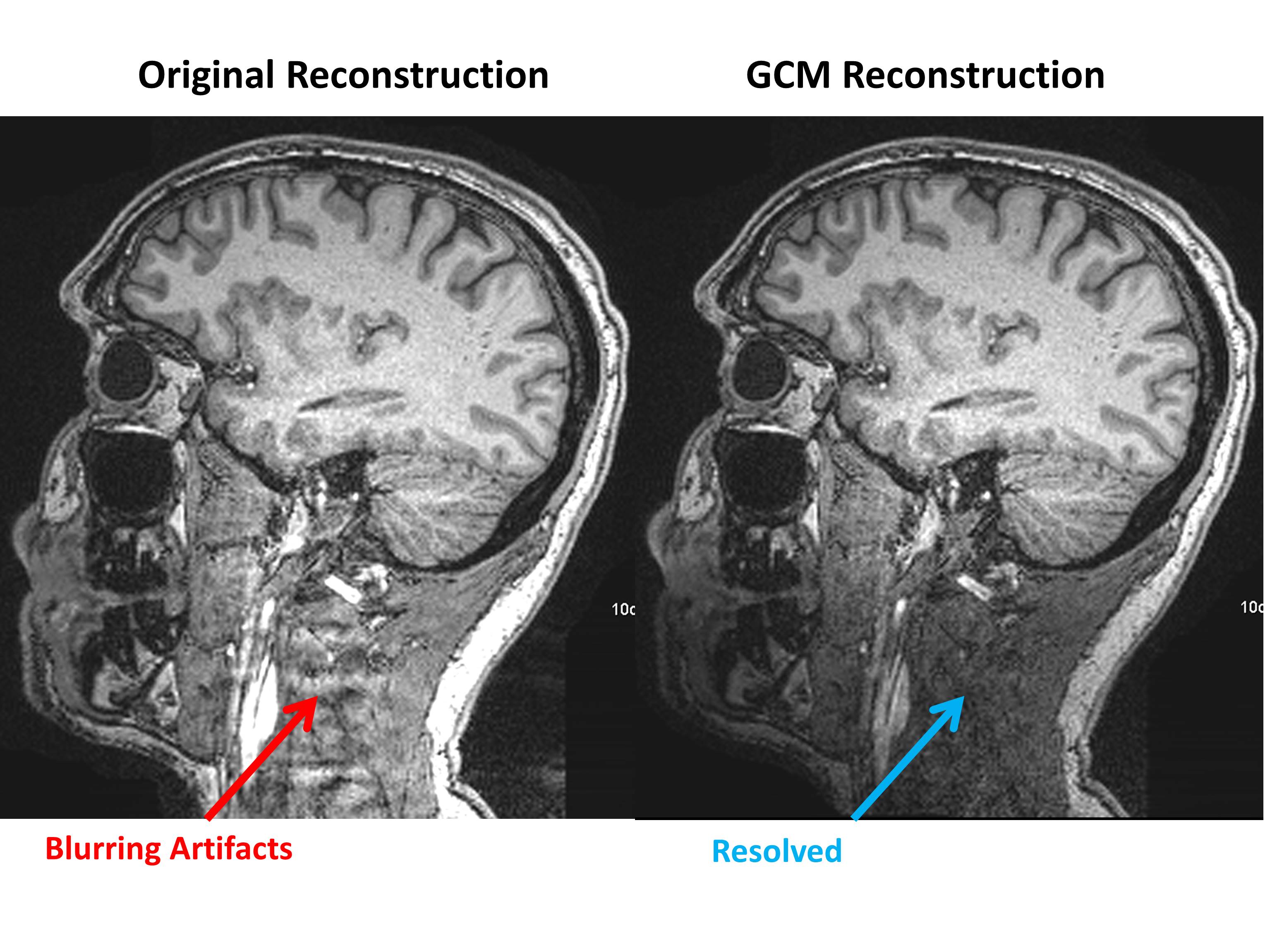
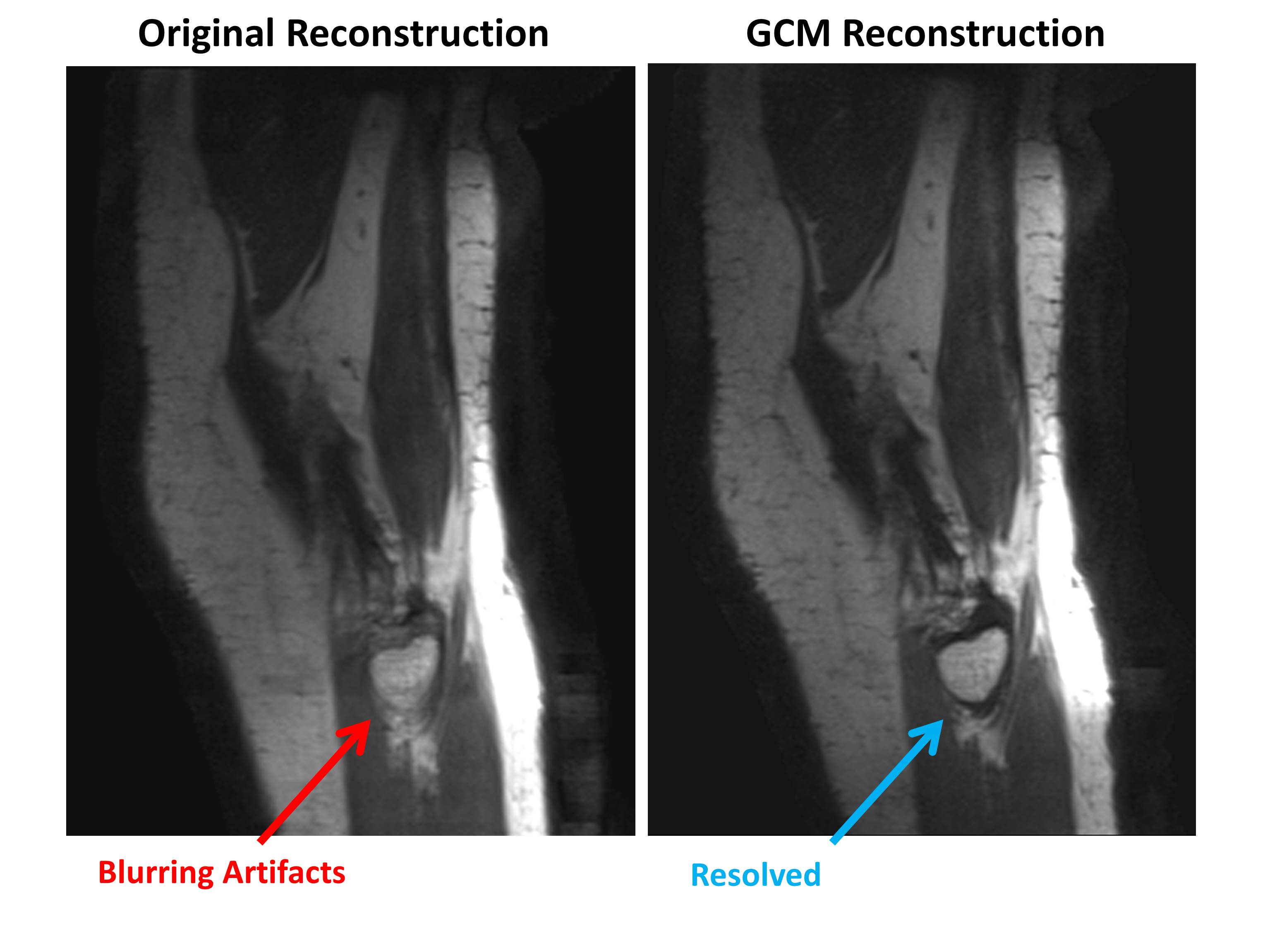
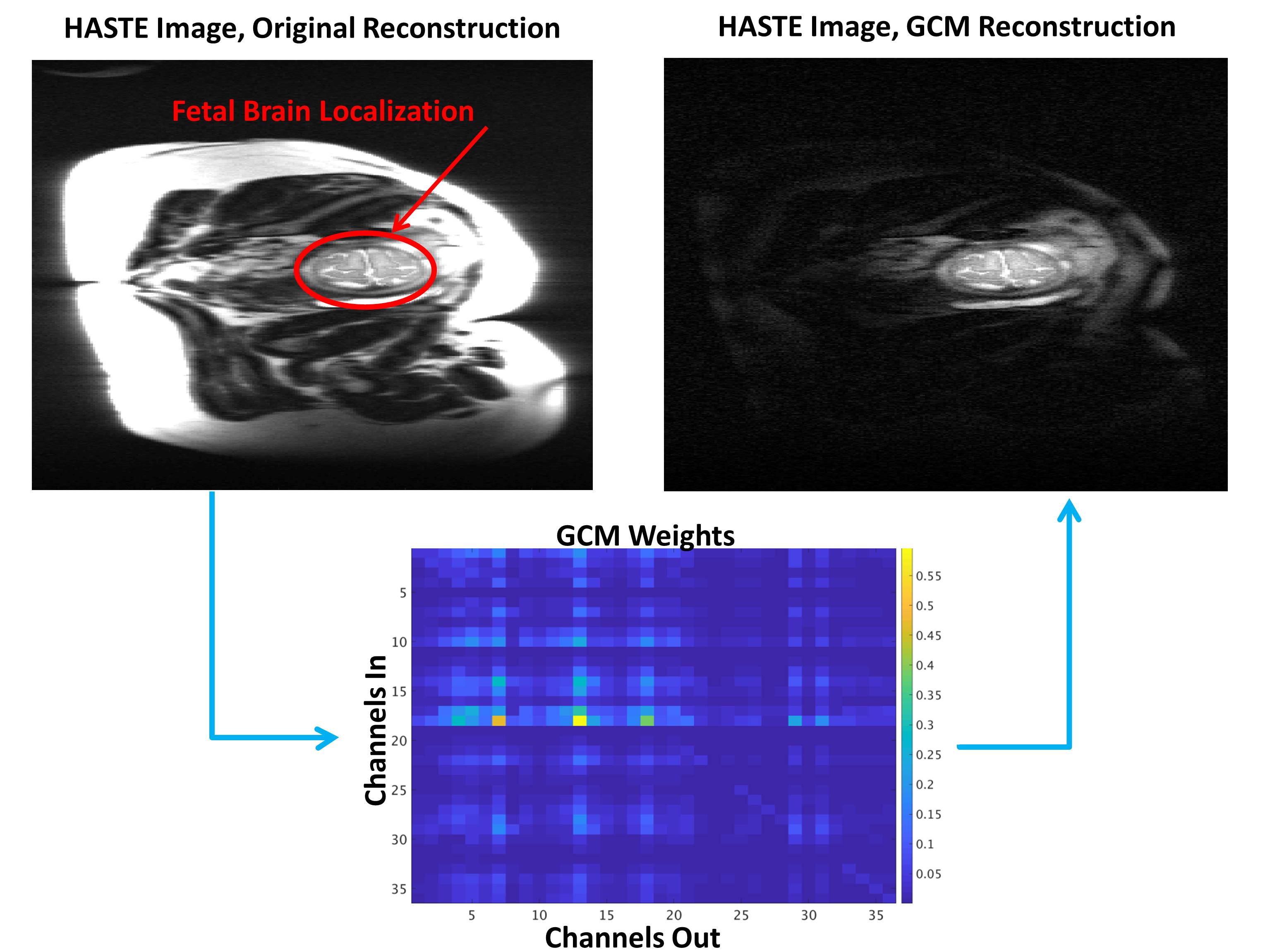
Fig.1 shows an illustration for the removal of shoulder signal that wraps into the FOVPAR during Wave-CAIPI acquisitions. Accurately separating the wrapped signal is difficult and any inaccuracies will lead to severe artifacts due to the wave encoding that produces aliasing along all spatial dimensions. Using the proposed GCM, we can restrict the signal sensitivity to a region that only contains the brain. Note that the resulting mixing is non-diagonal. Fig. 2 shows the benefits of this approach using a 64ch head/neck coil where high signal from the neck and shoulders creates artifacts that are likely to propagate throughout the brain based upon the R=3×3 readout-coupled Wave-CAIPI aliasing pattern. The original (left) and GCM (right) reconstructions are shown. GCM produced strong signal decay below the brain, which resulted in artifact reduction. Related simulations of gfactor showed only slight increases (~0.002) in the region of interest. Fig. 3 shows the presence of artifacts when applying the Wave-CAIPI SPACE sequence to image a knee. These artifacts are caused by non-modeled signal outside of the reconstructed FOV. By limiting the targeted receive region to within +/-10% of the FOV boundary we are able to dampen the signal sufficiently to mitigate these artifacts. The potential application of GCM to fetal imaging is illustrated in Fig. 4. A targeted receive region was specified around the fetal brain and the change in sensitivity profile for a standard clinical HASTE acquisition is shown. This mixing strategy has the potential to dampen the influence of non-rigid fetal and maternal motion, which can be problematic as the signal outside the fetal brain will change throughout the acquisition. It can also be incorporated into retrospective rigid-body motion correction techniques14,15 to improve the data consistency metric and further reduce artifacts from these non-rigid motion sources.
Discussion and Conclusion
We have demonstrated the utility of GCM for reducing artifacts caused by confounding signals within PI reconstruction. This situation-specific mixing can be determined efficiently through a least squares formulation using readily available auto-calibration data. Defining the targeted receive region allows for a trade-off between removing unwanted signal and retaining sensitivity (and related g-factor) across the region of interest. The potential incorporation of this technique into retrospective motion correction algorithms has been discussed for fetal brain imaging. Adjusting the GCM for each shot of data should allow for further improvement of the data consistency metric by dampening signals that do not fit the rigid-body motion model.Acknowledgements
This work was supported in part by NIH research grants: R01EB020613, U01HD087211, R01EB006847, R01EB019437, R24MH106096, P41EB015896, and the shared instrumentation grants: S10RR023401, S10RR019307, S10RR019254, S10RR023043References
1. Pruessmann, K. P., Weiger, M., Scheidegger, M. B. & Boesiger, P. SENSE: Sensitivity encoding for fast MRI. Magn. Reson. Med. 42, 952–962 (1999).
2. Sodickson, D. K. & Manning, W. J. Simultaneous acquisition of spatial harmonics (SMASH): Fast imaging with radiofrequency coil arrays. Magn. Reson. Med. 38, 591–603 (1997).
3. Griswold, M. A., Jakob, P. M., Nittka, M., Goldfarb, J. W. & Haase, A. Partially Parallel Imaging with Localized Sensitivities (PILS). Magn. Reson. Med. 44, 602–609 (2000).
4. Griswold, M. A. et al. Generalized Autocalibrating Partially Parallel Acquisitions (GRAPPA). Magn. Reson. Med. 47, 1202–1210 (2002).
5. Lustig, M., Donoho, D. & Pauly, J. M. Sparse MRI: The application of compressed sensing for rapid MR imaging. Magn. Reson. Med. 58, 1182–1195 (2007).
6. Liang, D., Liu, B., Wang, J. & Ying, L. Accelerating SENSE using compressed sensing. Magn. Reson. Med. 62, 1574–1584 (2009).
7. Roemer, P. B., Edelstein, W. A., Hayes, C. E., Souza, S. P. & Mueller, O. M. The NMR phased array. Magn. Reson. Med. 16, 192–225 (1990).
8. Keil, B. et al. A 64-channel 3T array coil for accelerated brain MRI. Magn. Reson. Med. 70, 248–258 (2013).
9. Buehrer, M., Pruessmann, K. P., Boesiger, P. & Kozerke, S. Array compression for MRI with large coil arrays. Magn. Reson. Med. 57, 1131–1139 (2007).
10. Huang, F., Vijayakumar, S., Li, Y., Hertel, S. & Duensing, G. R. A software channel compression technique for faster reconstruction with many channels. Magn. Reson. Imaging 26, 133–141 (2008).
11. King, S. B., Varosi, S. M. & Duensing, G. R. Optimum SNR data compression in hardware using an Eigencoil array. Magn. Reson. Med. 63, 1346–1356 (2010).
12. Zhang, T., Pauly, J. M., Vasanawala, S. S. & Lustig, M. Coil compression for accelerated imaging with Cartesian sampling. Magn. Reson. Med. 69, 571–582 (2013).
13. Bilgic, B. et al. Wave-CAIPI for highly accelerated 3D imaging. Magn. Reson. Med. 73, 2152–2162 (2015).
14. Cordero-Grande, L., Hughes, E. J., Hutter, J., Price, A. N. & Hajnal, J. V. Three-dimensional motion corrected sensitivity encoding reconstruction for multi-shot multi-slice MRI: Application to neonatal brain imaging. Magnetic Resonance in Medicine 0, (2017).
15. Haskell, M. W., Cauley, S. F. & Wald, L. L. TArgeted Motion Estimation and Reduction (TAMER): Data Consistency Based Motion Mitigation for MRI using a Reduced Model Joint Optimization. IEEE Trans. Med. Imaging (2018). doi:10.1109/TMI.2018.2791482
Figures