Cerebrovascular Vessel Wall Imaging from a Clinical Perspective
1University Medical Center Utrecht, Netherlands
Synopsis
Vessel wall MRI of the supra-aortic and intracranial vasculature has seen an exponential increase in popularity in the last two decades. It can provide a wealth of information on pathologic processes of the vessel wall associated with cerebrovascular diseases – like atherosclerosis, vasculitis, Moyamoya disease and aneurysms – that may be used in the differential diagnosis of vasculopathies, in (stroke) risk assessment, and for planning individual patient-based treatment strategies. This lecture will discuss the clinical potential of vessel wall MRI in these cerebrovascular diseases, with a specific focus on intracranial vessel wall pathology.
Background
Cerebrovascular diseases are associated with a high morbidity and mortality worldwide, and comprise a variety of common (e.g. atherosclerosis, intracranial aneurysms) and more rare (e.g. vasculitis, Moyamoya disease) pathologic processes of the supra-aortic extracranial and intracranial arteries. Imaging of cerebrovascular diseases can roughly be divided into 1. Visualizing their effect, i.e. ischemia, intracranial hemorrhage; and 2. Visualizing the causative pathologic process (vasculopathy). Although effect assessment – for instance ruling out hemorrhage so that intravenous thrombolytic agents can be infused as soon as possible – is critical in the acute phase, defining the causative pathologic process is essential for further treatment planning and prognosis. CT(A), MR(A) and DSA have traditionally been the mainstay for imaging both the causative vasculopathy and its effect. However, these techniques only visualize the vascular lumen instead of the pathologic vessel wall, limiting their diagnostic value. In the last two decades, focus has therefore shifted towards vessel wall imaging, which was initially restricted to the supra-aortic extracranial arteries but has more recently been extended to include the intracranial vasculature as well. The previous lecture by dr. Fan illustrated the technical considerations for supra-aortic vessel wall MRI; this lecture will describe their use in clinical decision-making, with a specific focus on the intracranial vasculature.Clinical applications – Atherosclerosis
Atherosclerosis is one of the most common vasculopathies of the supra-aortic extracranial and intracranial vasculature. The unstable, ‘vulnerable’ atherosclerotic plaque is the main cause of symptomatology, and is characterized by a thin fibrous cap, large lipid-rich necrotic core and intraplaque hemorrhage. Atherosclerotic plaques in the extracranial carotid artery (and vertebral artery to a lesser extent) have been studied with a diverse palette of specially developed MRI sequences all targeted to visualize these different plaque components. Although these sequences have not been implemented in current clinical guidelines yet – instead, arterial stenosis has remained the primary metric for assessing stroke risk and treatment planning – they can aid in identifying atherosclerotic plaques that have a higher risk of subsequent ischemic events.
Atherosclerosis of the intracranial arteries (ICAS) has only gained attention in the last decade after the realization that it is one of the main causes of ischemic stroke worldwide. ICAS’ intracranial phenotype – in contrast to its extracranial counterpart – seems to depend highly on ethnicity: patients from Asian descent generally show large atherosclerotic plaques that can be characterized similarly to atherosclerotic plaques in the extracranial carotid arteries, while Western Caucasian patients show smaller vessel wall lesions that are less readily characterized. In the latter case, plaque characterization has been attempted, but is limited due to the spatial resolution requirements as well as the lack of histologic validation (see below). Instead, lesion shape and enhancement pattern have been used to differentiate atherosclerosis from other non-atherosclerotic vasculopathies. Intracranial atherosclerotic lesions are characterized by their focal nature and eccentric shape, may or may not enhance, and can be found throughout the intracranial vasculature.
Clinical applications – Non-atherosclerotic vasculopathies & aneurysms
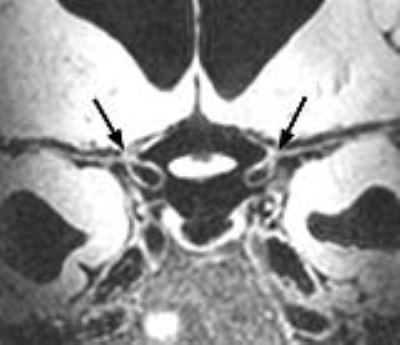
In addition to atherosclerosis, some other, much less common vasculopathies can be identified in the extracranial supra-aortic arteries, including Takayasu’s arteritis, fibromuscular dysplasia and arterial dissection. However, they are either associated with additional symptoms facilitating differentiation between these entities, or can be readily identified using lumenographic techniques. In the intracranial vasculature, major differential diagnoses are vasculitis, reversible cerebral vasoconstriction syndrome (RCVS) and Moyamoya disease. Clinically, these vasculopathies can be difficult to differentiate from one another, especially vasculitis versus atherosclerosis and RCVS; however, treatment of these entities is completely different. Vessel wall MRI can aid in differentiating between these vasculopathies, showing more concentric, diffuse vessel wall thickening throughout the intracranial vasculature in vasculitis, and vessel wall abnormalities restricted to the distal internal carotid artery and proximal middle cerebral artery in Moyamoya disease.
Intracranial aneurysms can easily be identified using lumenographic techniques; nonetheless, it has proven difficult to predict their risk of rupture using only their luminal size and shape. Studies visualizing the aneurysm wall have shown that wall enhancement may be a marker for rupture risk, while wall thickness is inversely correlated with wall shear stress. However, these are still preliminary results in relatively small study groups compared to the data available for general vessel wall imaging.
Considerations for clinical implementation
The primary difference between intracranial arteries and their extracranial counterparts is that validation of MRI results is difficult if not impossible for the intracranial vasculature: there is no such thing as e.g. ‘middle cerebral artery endarterectomy’, and consequently no opportunity for in vivo - ex vivo comparison. Pathology studies are restricted to postmortem material with all its drawbacks. This also means that results from studies utilizing intracranial vessel wall imaging need to be interpreted with caution, especially when used in clinical practice. For instance, we do not really know what tissue enhances when we observe an enhancing vessel wall lesion; we assume it might be inflammation or vasa vasorum, but we do not know this to be true. Furthermore, although already largely defined for the extracranial supra-aortic arteries, necessary spatial resolution, image contrast weightings and magnetic field strength have not yet been established for intracranial vessel wall MRI. Finally, the added value of these vessel wall imaging techniques in clinical decision-making and final outcome has not been addressed yet, and will be necessary for further clinical implementation.Conclusion
Vessel wall MRI of the supra-aortic extracranial and intracranial arteries provides a wealth of information on pathologic processes of the vessel wall associated with cerebrovascular diseases. Image characteristics of vessel wall pathology may be used for (stroke) risk assessment, and could enable differentiation between vasculopathies, thereby improving individual patient-based treatment strategies.Acknowledgements
No acknowledgement found.References
- Saba L, Yuan C, Hatsukami TS, et al. Carotid Artery Wall Imaging: Perspective and Guidelines from the ASNR Vessel Wall Imaging Study Group and Expert Consensus Recommendations of the American Society of Neuroradiology. AJNR 2018; doi: 10.3174/ajnr.A5488 [Epub ahead of print]
Recent perspective article including guidelines on vessel wall MRI of the extracranial carotid artery
- Lindenholz A, van der Kolk AG, Zwanenburg JJM, Hendrikse J. The Use and Pitfalls of Intracranial Vessel Wall Imaging: How We Do It. Radiology 2018; 286: 12-28
Recent article discussing technical requirements for intracranial vessel wall MRI, as well as step-by-step clinical assessment of vessel wall images
- Mandell DM, Mossa-Basha M, Qiao Y, et al. Intracranial Vessel Wall MRI: Principles and Expert Consensus Recommendations of the American Society of Neuroradiology. AJNR 2017; 38: 218-229
Perspective article including recommendations on vessel wall MRI of the intracranial vasculature
- Ritz K, Denswil NP, Stam OCG, van Lieshout JJ, Daemen MJAP. Cause and Mechanisms of Intracranial Atherosclerosis. Circulation 2014; 130: 1407-1414
Provides an overview of etiology, morphology and risk factors of intracranial atherosclerosis
- Lehman VT, Brinjikji W, Mossa-Basha M, et al. Conventional and high-resolution vessel wall MRI of intracranial aneurysms: current concepts and new horizons. J Neurosurg 2017; doi: 10.3171/2016.12.JNS162262 [Epub ahead of print]
Review article of recent literature on vessel wall MRI of the intracranial aneurysm wall
Figures
