Clinical Translation & Applications of Molecular MRI in Cardiovascular Diseases
1King's College London, United Kingdom
Synopsis
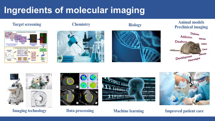
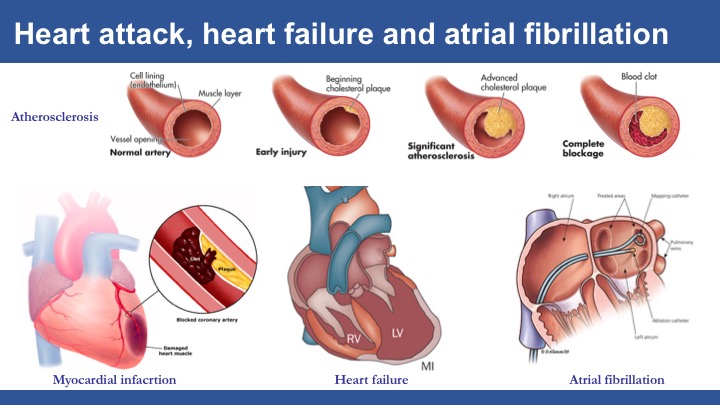
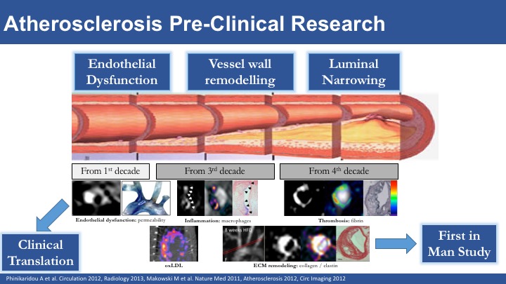
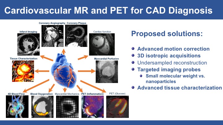
Molecular magnetic resonance imaging (MRI) is a promising tool to detect molecular and cellular changes in the carotid, aortic and coronary vessel wall including endothelial dysfunction, inflammation, vascular remodelling, enzymatic activity, intraplaque haemorrhage and fibrin deposition and thus may allow early detection of unstable lesions and improve the prediction of future coronary events. To increase the biological information obtained by MRI a variety of targeted-specific molecular probes have been developed for the non-invasive visualisation of particular biological processes at the molecular and cellular level. This presentation will discuss recent advances in molecular MRI of atherosclerosis, covering both pulse sequence development and also the design of novel contrast agents, for imaging atherosclerotic disease in vivo.
Acknowledgements
No acknowledgement found.References
1. Abubakar I, Tillmann T, Banerjee A (2015) Global, regional, and national age-sex specific all-cause and cause-specific mortality for 240 causes of death, 1990-2013: a systematic analysis for the Global Burden of Disease Study 2013. Lancet 385:117-171 doi:10.1016/S0140-6736(14)61682-2
2. Weber, C. & Noels, H (2011) Atherosclerosis: current pathogenesis and therapeutic options. Nature medicine 17, 1410-1422, doi:10.1038/nm.2538 .
3. Stone, G. W. et al (2011) A prospective natural-history study of coronary atherosclerosis. The New England journal of medicine 364, 226-235, doi:10.1056/NEJMoa1002358
4. Caravan, P., Ellison, J. J., McMurry, T. J. & Lauffer, R. B (1999) Gadolinium(III) Chelates as MRI Contrast Agents: Structure, Dynamics, and Applications. Chemical reviews 99, 2293-2352
5. Kooi, M. E. et al (2003) Accumulation of ultrasmall superparamagnetic particles of iron oxide in human atherosclerotic plaques can be detected by in vivo magnetic resonance imaging. Circulation 107, 2453-2458, doi:10.1161/01.CIR.0000068315.98705.CC
6. Lancelot, E. et al (2008) Evaluation of matrix metalloproteinases in atherosclerosis using a novel noninvasive imaging approach. Arteriosclerosis, thrombosis, and vascular biology 28, 425-432, doi:10.1161/ATVBAHA.107.149666
7. Makowski, M. R. et al (2011) Assessment of atherosclerotic plaque burden with an elastin-specific magnetic resonance contrast agent. Nature medicine 17, 383-388, doi:10.1038/nm.2310
8. Phinikaridou, A. et al (2012) Noninvasive magnetic resonance imaging evaluation of endothelial permeability in murine atherosclerosis using an albumin-binding contrast agent. Circulation 126, 707-719, doi:10.1161/CIRCULATIONAHA.112.092098
9. Noguchi, T. et al (2014) High-intensity signals in coronary plaques on noncontrast T1-weighted magnetic resonance imaging as a novel determinant of coronary events. Journal of the American College of Cardiology 63, 989-999, doi:10.1016/j.jacc.2013.11.034
Figures