An introduction to Synthetic MRI
Marcel Warntjes1,2
1CMIV, Linköping University, Sweden, 2SyntheticMR, Linköping, Sweden
Synopsis
Synthetic Magnetic Resonance Imaging has been around since the early days of MRI, but only recently the acquisition and post-processing have become fast enough to apply it clinically. In essence, it is a rapid quantification of MRI properties and a subsequent recreation, or synthesis, of conventional images.
Summary
Synthetic Magnetic Resonance Imaging has been around since the early days of MRI, but only recently the acquisition and post-processing have become fast enough to apply it clinically. In essence, it is a rapid quantification of MRI properties and a subsequent recreation, or synthesis, of conventional images. The difference with conventional imaging therefore, becomes the freedom to adjust scanner parameters after the patient has left. But since it is based on quantitative MRI, much more can be done, for example automatic tissue recognition. The technique, history, clinical application and pitfalls of synthetic MRI will be covered. The approach can provide a wealth of clinical information within a short scan time and many examples will be shown.Acknowledgements
No acknowledgement found.References
No reference found.Figures
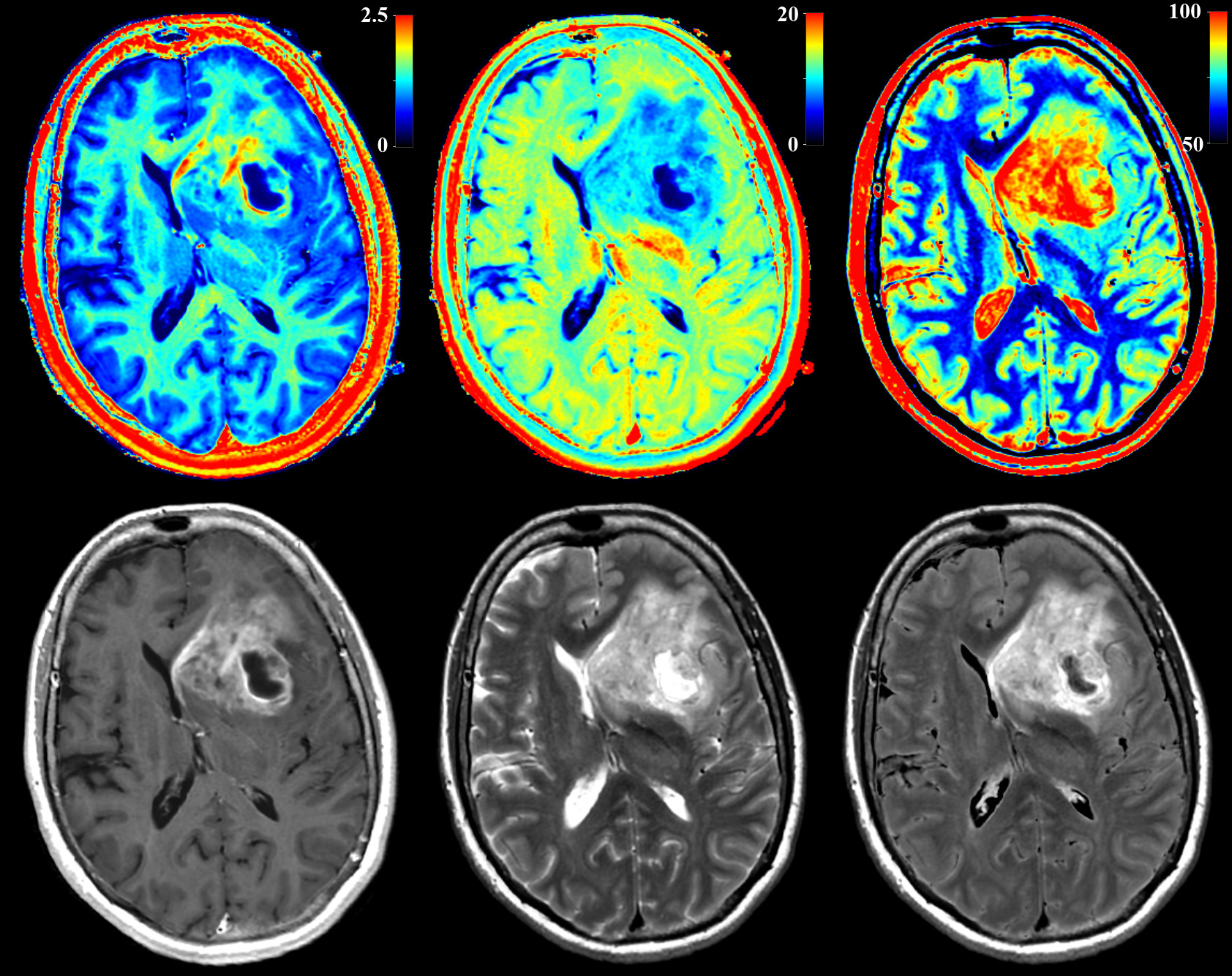
Example of synthetic MRI: At first, the MR properties of R1 relaxation rate, R2 relaxation rate and proton density PD are measured. An axial slice of a glioma patient is displayed in the top row, with R1 on a scale 0-2.5s-1, R2 on a scale 0-20s-1 and PD on a scale 50-100%. Using R1, R2 and PD, conventional images such as T1W, T2W and FLAIR can be synthesized (bottom row). All images were acquired in a single sequence of 6 minutes.