Image Quality
1Division of Radiological Physics, Dept. of Radiology, University Hospital Basel, Basel, Switzerland, 2Dept. of Biomedical Engineering, University of Basel, Basel, Switzerland
Synopsis
This educational talk presents an overview of key aspects influencing and quantitatively depicting image quality. Several examples for the different types of image quality aspects are given. The importance of signal-to-noise ratio (SNR) and the "triangle of death" for image quality in MRI is discussed in particular.
Motivation
Magnetic Resonance Imaging (MRI) has experienced tremendous growth since its establishment in the clinical practice in the early 1980s. For the successful application as a medical imaging modality, however, the resulting image quality is of utmost importance for MRI’s diagnostic accuracy.
Numerous factors influence the MR image quality. For this reason, improving the quality of a given MRI acquisition can be challenging sometimes; besides, defining MR image quality can be rather difficult.
Commonly, a “good” or “valuable MRI protocol” represents a trade-off between different image quality aspects. It is always optimized for dedicated organs and pathologies.
The image quality aspects can be classified into three categories:
- Resolution, volume coverage, k-space sampling, and signal-to-noise-ratio (SNR)
- Contrast generation
- Artifacts
Resolution, volume coverage, k-space sampling, and SNR
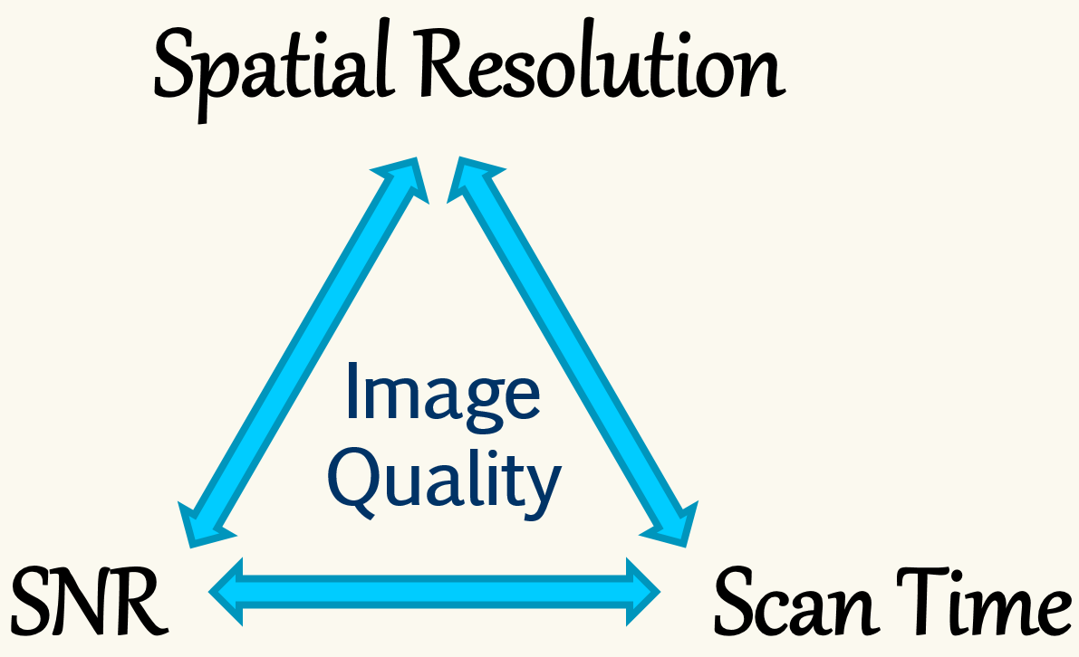
Protocol parameter changes affecting the resolution, volume coverage, or k-space sampling usually have an impact on the SNR. Typically, an increase of image quality can only be achieved at the cost (i.e. with a reduction) of SNR. Since MRI has generally a low sensitivity, the importance of SNR cannot be overestimated in MRI.
Generally, based on physics laws, it can be shown that the SNR of an MRI acquisition is proportional to the voxel size ΔV (-> resolution) and to the square root of the total acquisition time TA: SNR ~ ΔV × √TA.
Spatial encoding is rather time-consuming in MRI. So, increasing the resolution is generally time- and SNR-intensive.
For a desired increase of SNR, the user frequently invests more scan time (see above), which is already rather long for MRI compared to other imaging modalities. Overall, the adverse interdependency of resolution, SNR, and scan time leads to the "triangle of death" in MR image quality as outlined in Fig. 1.
Contrast generation
Tissue contrast in the image – that is essential for diagnosis - is mainly generated by the various MRI sequences and preparation modules, harnessing the different physical MR tissue properties such as proton density PD, relaxation times T1 and T2, diffusion, magnetization transfer (MT), susceptibility, or flow. Furthermore, several MR tissue properties change with pathological conditions; some of these are also field strength dependent.
Typical MRI contrast parameters like echo time TE, repetition time TR, inversion time TI, or flip angle FA, control the impact of these MR tissue properties on the final image contrast.
Artifacts
Artifacts represent structures in the image that do not reflect the “real tissue morphology” in the imaged body. In order to avoid misinterpretations and wrong diagnoses, it is important to spot and identify artifacts.
Based on their primary source, artifacts can roughly be classified into three sub categories:
- Physical artifacts (basic NMR effect, MR imaging principle)
- Technical artifacts that are MR system – related
- Physiological artifacts, generated by the living body
Though - it is to say - that particularly the categories of physical and technical artifacts can often not be completely separated, they do overlap for some artifacts.
As a final note, it should be mentioned that patient safety restrictions such as the specific absorption rate (SAR) or peripheral nerve stimulation (PNS) can have an indirect, adverse effect on image quality. Money may be another factor (MR system hardware and software).
This educational talk presents an overview of key aspects influencing and quantitatively depicting image quality. Several examples for the different types of image quality aspects are given.
Acknowledgements
No acknowledgement found.References
No reference found.Figures