Hyperpolarized 13C MRI: The Technical Aspects from Hamiltonian to Homo sapien
1University of Oxford, United Kingdom
Synopsis
We will examine the mechanistic details behind dissolution-Dynamic Nuclear Polarisation with small molecules for in vivo applications, and additionally discuss some of the technical challenges that arise in the design of pulse sequences to use the magnetisation that the technique generates.
Precis
Magnetic Resonance techniques are traditionally viewed as being inherently insensitive. This insensitivity arises due to the Boltzmann equation: at temperatures compatible with life and at plausible field strengths, the limiting nuclear polarization that can be obtained is on the order of $$$P\approx10^{-6}$$$. This serves to frustrate many potential applications of magnetic resonance, particularly 13C spectroscopy performed on highly biologically relevant small molecules present at physiologically small concentrations.
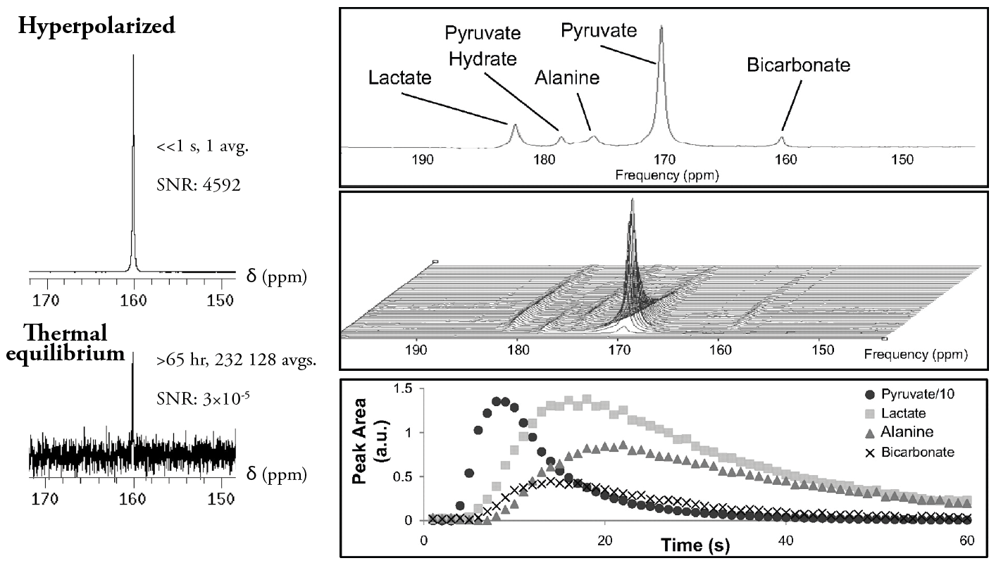
Hyperpolarization methods aim to overcome the fundamental limiting polarization by using additional physical means to create a large nuclear polarization exogenously, prior to the preparation and reintroduction of the out-of-thermodynamic equilibrium sample to the detection or imaging magnet. The large magnetisation transiently generated by the hyperpolarization method employed creates a large NMR signal, typically three to five orders of magnitude larger than that expected thermally. The magnetisation generated then decays exponentially to thermal equilibrium, with its $$$T_1$$$.
This talk will briefly examine the mechanistic basis and applications for the one hyperpolarization technique that has entered clinical trials, dissolution Dynamic Nuclear Polarization (d-DNP). Dynamic Nuclear Polarization functions by mixing a labelled compound of interest (e.g. [1-$$$^{13}$$$C]pyruvic acid for human applications) together with a source of electronic free radicals present in small concentrations, freezing the resulting liquid as a glass, and placing it in avariable-temperature modified NMR system, commonly at $$$B\approx3.35$$$ or $$$5$$$ Tesla, at approximately 1 K. Under these conditions, essentially all electronic spins are thermally in their ground state, but the Boltzmann distribution over nuclear spins is still approximately 50-50. Subsequent continuous irradiation with photons of the appropriate (microwave) energy can therefore promote the enhancement of nuclear polarization through a variety of microscopic mechanisms, known as the solid effect,the cross effect, and thermal mixing. After nuclear polarization has been generated to a limiting value (commonly $$$P\sim0.5\%$$$), the sample can be rapidly melted through the introduction of a superheated solvent at $$$\sim10^6$$$ Pa and $$$\sim200\,\,^{\circ}$$$C. The resulting liquid has a high polarization and a $$$T_1$$$ that is typically on the order of tens of seconds [1]. It can then be rapidly injected into a biological system under study in a nearby imaging magnet, and followed through its subsequent biochemistry over time and in three-dimensional space [2] (c.f. Fig 1).
The use of d-DNP to hyperpolarize small metabolites such as pyruvate has enabled a new field of metabolic imaging. The ordinarily minute thermal equilibrium 13C NMR signal ensures that all hyperpolarized experiments have essentially no background, and provide a clear window on to the ultimate bio-distribution of the injected agent. The finite and non-renewable pool of magnetisation generated by d-DNP creates several challenges for the pulse sequence programmer, as the imaging of hyperpolarized compounds necessitates minimising the total amount of RF excitation while still achieving conflicting goals of high temporal, spatial and chemical resolution in a dynamic timecourse. We will explore these challenges, and briefly survey both the future promise of d-DNP as a clinical tool and the large number of important scientific results to which it has already contributed.
Acknowledgements
I would like to acknowledge financial support from the British Heart Foundation, Novo Nordisk, and the Engineering and Physical Sciences Research Council, and thank ISMRM for the opportunity to present.References
[1] J. H. Ardenkjaer-Larsen, B. Fridlund, A. Gram, G. Hansson, L. Hansson, M. H. Lerche, R. Servin, M. Thaning, and K. Golman, “Increase in signal-to-noise ratio of > 10,000 times in liquid-state NMR.,” Proc. Natl. Acad. Sci. U. S. A., vol. 100, no. 18, pp. 10158–63, Sep. 2003.
[2] K. Golman, J. H. Ardenkjaer-Larsen, J. S. Petersson, S. Mansson, and I. Leunbach, “Molecular imaging with endogenous substances.,” Proc. Natl. Acad. Sci. U. S. A., vol. 100, no. 18, pp. 10435–9, Sep. 2003.
Figures