Correcting Cardiac Motion
1Biomedical Engineering, Illinois Institute of Technology, Chicago, IL, United States, 2Medicine, The University of Chicago, Chicago, IL, United States
Synopsis
Early efforts towards the correction for cardiac motion have focused on addressing respiratory motion of the heart, and particularly for imaging of the coronary arteries. After over two decades of key developments that include navigator gating, self-navigation, and more elaborate 2D and 3D variants of these navigator approaches, numerous motion correction methods have proliferated to almost every aspects of cardiac MRI, including LGE, cine, relaxometry mapping approaches, tagging, perfusion, and new inherently 3D methods such as Quantitative Susceptibility Mapping. This presentation will additionally cover the current state-of-the-art methods that include highly advanced approaches in cardiovascular MRI research today.
Highlights
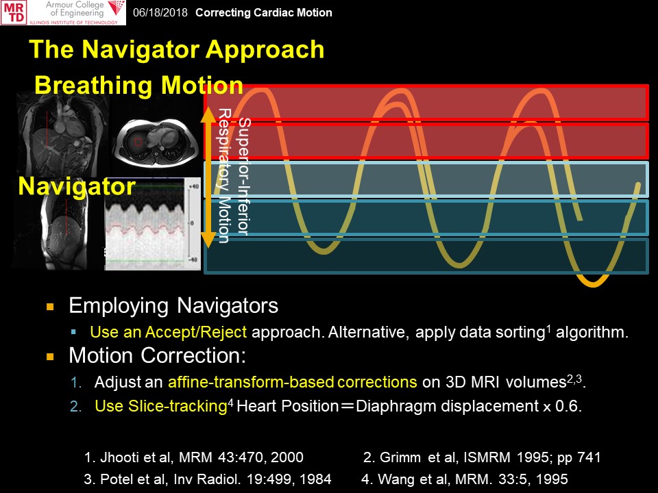
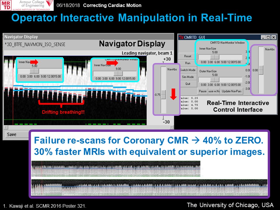
1) Navigator approaches as either separate echo acquisitions or self-gated acquisitions can provide key physiologic information to overcome respiratory motion that leads to notable displacement of the heart.
2) New cardiac motion correction approaches that are tailored to specific Cardiac MR assessments may offer improved evaluation of clinical evaluations of the heart, and add diagnostic value in MR-based assessment of cardiovascular diseases and conditions.
Target Audience
Scientists and clinicians with basic knowledge of MRI physics and pulse sequence design who either conduct research to address motion problems in MRI, or conduct MRI exams in the heart for clinical studies.Introduction
This lecture on correcting cardiac motion is divided into two parts.
In part 1, we will summarize the technical development and clinical translation process of numerous clinical and advanced research cardiac motion correction methods available today.
In part 2, we will provide examples of novel cardiac motion correction methods and techniques, and thereby provide a big-picture overview of where Cardiac MR (CMR) may lead to in the near future.
Part 1: The Navigator Approach and Respiratory Motion Correction – from initial efforts towards 3D Coronary Imaging to the Comprehensive CMR protocol.
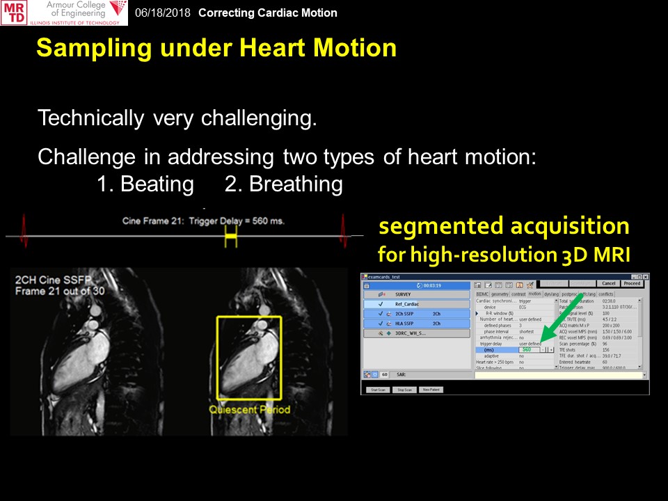
In this first part, we discuss prior works with cardiac motion correction that go hand-in-hand with the technical development of MRI-based methods to obtain high-resolution, motion-free images of the heart. Among these, imaging of the coronary arteries was considered among the most critical challenges over the past several decades, and notably while alternative modality approaches such as X-ray and CT often required significantly higher ionizing radiation dosages than what they require today.
Over this two-decade period, key research and developments were made towards achieving superior images of the coronary arteries in the heart through innovations in: a) image acquisition, b) real-time and post-processing, and c) systems instrumentation. Key features of these techniques are now widely available in the clinical MRI system settings, and these have helped establish the technical foundation of cardiac motion correction that are applicable to techniques beyond the originally intended high-resolution and motion-resolved coronary artery imaging.
Examples of how motion correction is embedded during non-conventional magnetization preparation phases of CMR pulse sequence designs for techniques including LGE, T1 mapping, 3D-Cine, and novel (and inherently 3D) approaches such as cardiac Quantitative Susceptibility Mapping, will be described.
Part 2: Emerging correction methods beyond overcoming motion artifacts for cardiac MRI.
In this second part, we discuss how the accumulated knowledge pool from prior efforts over several decades have evolved into today’s state-of-the-art cardiac imaging using MRI – both in clinical and pre-clinical research space. Select contribution slides by key innovators in this field will be provided to offer a broad picture of how uniquely tailored motion correction techniques may address specific cardiac MRI challenges. There will be specific focus as the cardiac MRI field transitions from qualitative assessment to more quantitative assessment methods that report direct measurements in absolute terms. Key topics for this section include:
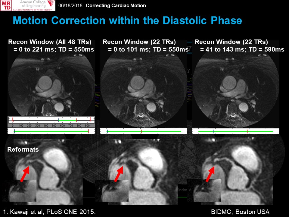
- Motion correction within the cardiac phase, and expanding to free-breathing methods for increased scan utilization within the cardiac cycle (non-Cartesian methods and 4D/5D methods).
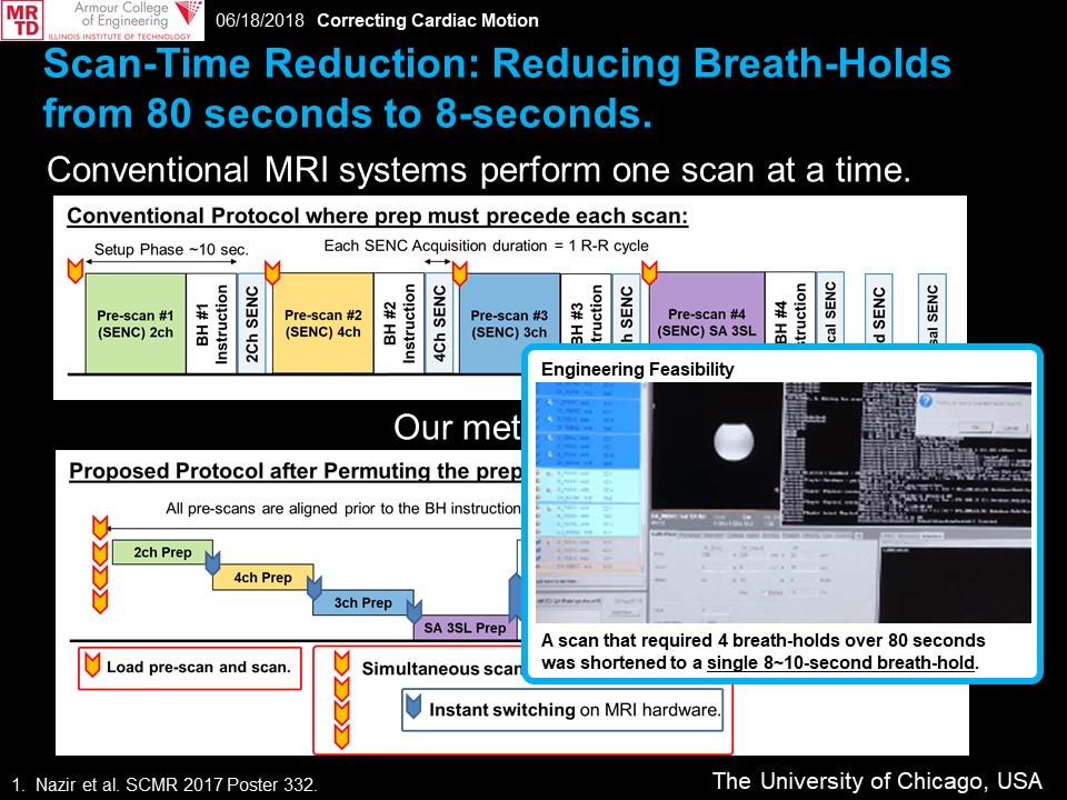
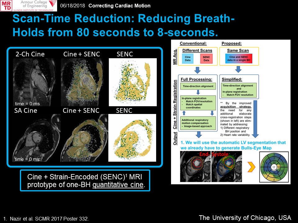
- Combined acquisition, motion correction and reconstruction strategies, notably for dynamic cardiac sequences (simultaneous acquisitions; perfusion, navigator-gated T1 mapping, etc.).
- Inter-related reconstruction/post-processing strategies to overcome either: imperfect breath-held or free-breathing motion in new CMR pulse sequences.
Acknowledgements
We thank all investigators who contributed research slides for this education presentation.
References
1. Wang Y, Riederer SJ, and Ehman RL. Respiratory motion of the heart: kinematics and the implications for the spatial resolution in coronary imaging. Magn Reson Med. 1995;33:713-719.
2. Stuber M, Botnar RM, Danias PG, Kissinger KV, and Manning WJ. Breathhold three-dimensional coronary magnetic resonance angiography using real-time navigator technology. J Cardiovasc Magn Reson. 1999;1(3):233-8.
3. Jhooti P, Gatehouse PD, Keegan J, Bunce NH, Taylor AM, Firmin DN. Phase ordering with automatic window selection (PAWS): a novel motion-resistant technique for 3D coronary imaging. Magn Reson Med. 2000;43(3):470-80. 4. Huber ME, Hengesbach D, Botnar RM, Kissinger KV, Boesiger P, Manning WJ, Stuber M. Motion artifact reduction and vessel enhancement for free-breathing navigator-gated coronary MRA using 3D k-space reordering. Magn Reson Med. 2001;45(4):645-52.
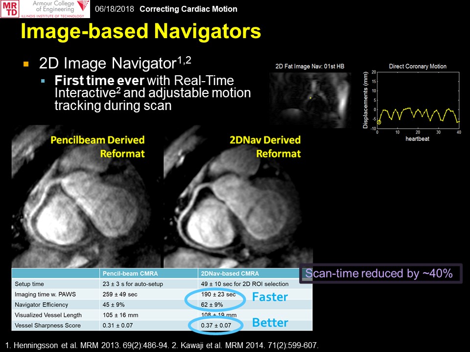
5. Henningsson M, Smink J, Razavi R, and Botnar RM. Prospective respiratory motion correction for coronary MR angiography using a 2D image navigator. Magn Reson Med. 2013;69(2):486-94.
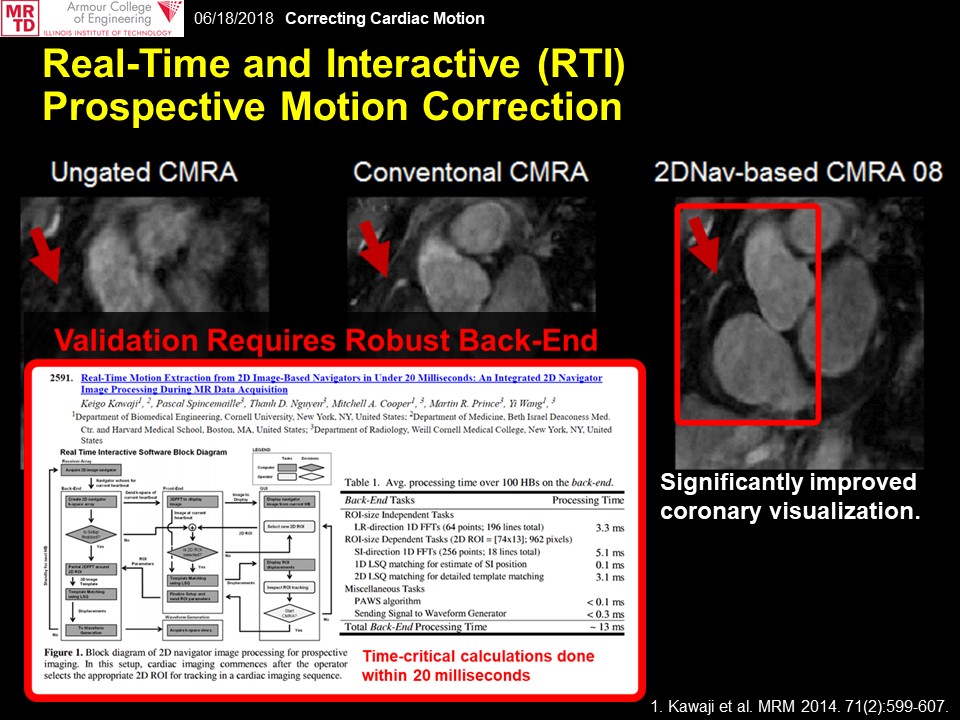
6. Kawaji K, Spincemaille P, Nguyen TD, Thimmappa N, Cooper MA, Prince MR, and Wang Y. Direct coronary motion extraction from a 2D fat image navigator for prospectively gated coronary MR angiography. Magn Reson Med. 2014;71(2):599-607.
7. Keegan J, Gatehouse PD, Yang GZ, Firmin DN. Non-model-based correction of respiratory motion using beat-to-beat 3D spiral fat-selective imaging. J Magn Reson Imaging. 2007;26(3):624-9.
8. Luo J, Addy NO, Ingle RR, Baron CA, Cheng JY, Hu BS, Nishimura DG. Nonrigid Motion Correction With 3D Image-Based Navigators for Coronary MR Angiography. Magn Reson Med. 2017;77(5):1884-1893.
9. Addy NO, Ingle RR, Luo J, Baron CA, Yang PC, Hu BS, Nishimura DG. 3D image-based navigators for coronary MR angiography. Magn Reson Med. 2017;77(5):1874-1883.
10. Moghari MH, Akçakaya M, O'Connor A, Basha TA, Casanova M, Stanton D, Goepfert L, Kissinger KV, Goddu B, Chuang ML, Tarokh V, Manning WJ, and Nezafat R. Compressed-sensing motion compensation (CosMo): a joint prospective-retrospective respiratory navigator for coronary MRI. Magn Reson Med. 2011;66(6):1674-81.
11. Moghari MH, Annese D, Geva T, Powell AJ. Three-dimensional heart locator and compressed sensing for whole-heart MR angiography. Magn Reson Med. 2016;75(5):2086-93.
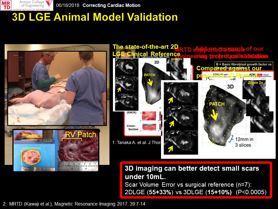
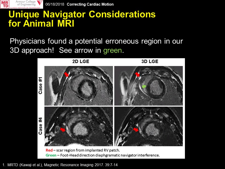
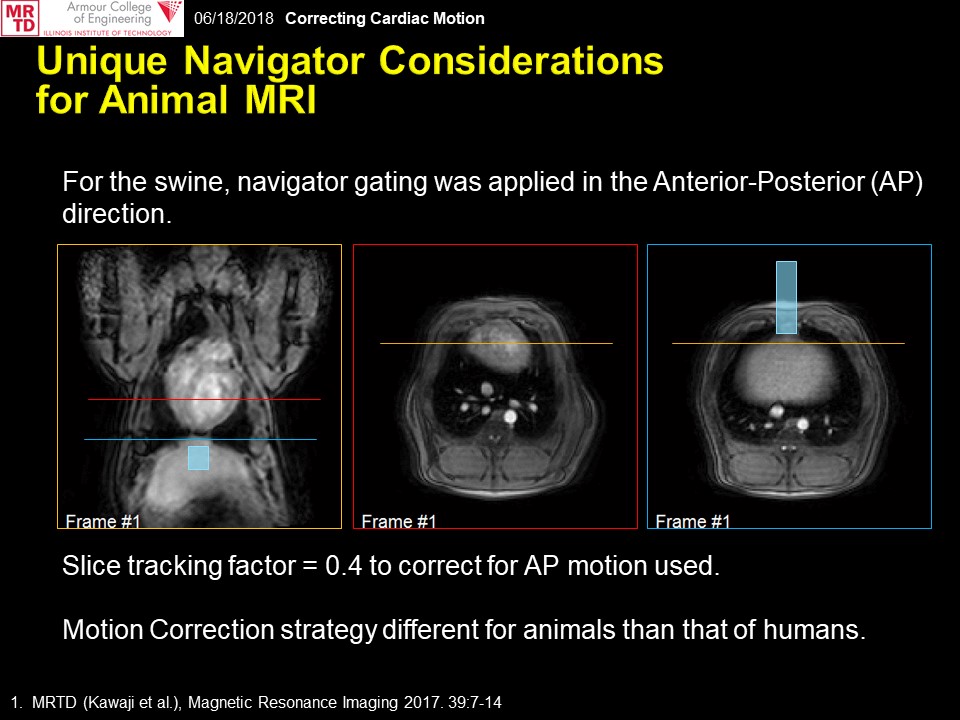
12. Kawaji K, Tanaka A, Patel MB, Wang H, Maffessanti F, Ota T, and Patel AR. 3D late gadolinium enhanced cardiovascular MR with CENTRA-PLUS profile/view ordering: Feasibility of right ventricular myocardial damage assessment using a swine animal model. Magn Reson Imaging. 2017;39:7-14.
13. Ibrahim el-SH, Stuber M, Fahmy AS, Abd-Elmoniem KZ, Sasano T, Abraham MR, Osman NF. Real-time MR imaging of myocardial regional function using strain-encoding (SENC) with tissue through-plane motion tracking. J Magn Reson Imaging. 2007;26(6):1461-70.
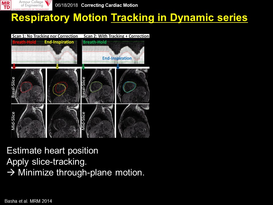
14. Basha TA, Roujol S, Kissinger KV, Goddu B, Berg S, Manning WJ, Nezafat R. Free-breathing cardiac MR stress perfusion with real-time slice tracking. Magn Reson Med. 2014;72(3):689-98.
15. Chow K, Yang Y, Shaw P, Kramer CM, Salerno M. Robust free-breathing SASHA T1 mapping with high-contrast image registration. J Cardiovasc Magn Reson. 2016 Aug 17;18(1):47.
16. Moghari MH, Barthur A, Amaral ME, Geva T, and Powell AJ. Free-breathing whole-heart 3D cine magnetic resonance imaging with prospective respiratory motion compensation. Magn Reson Med 2018;80(1):181-189.
17. Wen Y, Nguyen TD, Liu Z, Spincemaille P, Zhou D, Dimov A, Kee Y, Deh K, Kim J, Weinsaft JW, Wang Y. Cardiac quantitative susceptibility mapping (QSM) for heart chamber oxygenation. Magn Reson Med. 2018;79(3):1545-1552.
18. Han F, Zhou Z, Cao M, Yang Y, Sheng K, and Hu P. Respiratory motion-resolved, self-gated 4D-MRI using rotating cartesian k-space (ROCK). Med Phys. 2017;44(4):1359-1368.
19. Liu J and Saloner D. Accelerated MRI with CIRcular Cartesian UnderSampling (CIRCUS): a variable density Cartesian sampling strategy for compressed sensing and parallel imaging. Quant Imaging Med Surg. 2014;4(1):57-67.
20. Stehning C, Börnert P, Nehrke K, Eggers H, Stuber M. Free-breathing whole-heart coronary MRA with 3D radial SSFP and self-navigated image reconstruction. Magn Reson Med. 2005;54(2):476-80.
21. Feng L, Coppo S, Piccini D, Yerly J, Lim RP, Masci PG, Stuber M, Sodickson DK, Otazo R. 5D whole-heart sparse MRI. Magn Reson Med. 2018;79(2):826-838.
22. Pang J, Sharif B, Fan Z, Bi X, Arsanjani R, Berman DS, Li D. ECG and navigator-free four-dimensional whole-heart coronary MRA for simultaneous visualization of cardiac anatomy and function. Magn Reson Med. 2014;72(5):1208-17.
23. Peters DC, Nezafat R, Eggers H, Stehning C, and Manning WJ. 2D free-breathing dual navigator-gated cardiac function validated against the 2D breath-hold acquisition. J Magn Reson Imaging. 2008;28(3):773-7.
24. Hu C, Huber S, Latif SR, Santacana-Laffitte G, Mojibian HR, Baldassarre LA, Peters DC. Reverse double inversion-recovery: Improving motion robustness of cardiac T2 -weighted dark-blood turbo spin-echo sequence. J Magn Reson Imaging. 2017. In press. doi: 10.1002/jmri.25886
25. Harrison A, Adluru G, Damal K, Shaaban AM, Wilson B, Kim D, McGann C, Marrouche NF, DiBella EV. Rapid ungated myocardial perfusion cardiovascular magnetic resonance: preliminary diagnostic accuracy. J Cardiovasc Magn Reson. 2013 Mar 27;15:26.
26. Bonanno G, Hays AG, Weiss RG, Schär M. Self-gated golden angle spiral cine MRI for coronary endothelial function assessment. Magn Reson Med. 2017. In press. doi: 10.1002/mrm.27060
27. Krishnamurthy R, Pednekar A, Atweh LA, Vogelius E, Chu ZD, Zhang W, Maskatia S, Masand P, Morris SA, Krishnamurthy R, Muthupillai R. Clinical validation of free breathing respiratory triggered retrospectively cardiac gated cine balanced steady-state free precession cardiovascular magnetic resonance in sedated children. J Cardiovasc Magn Reson. 2015 Jan 14;17(1):1.
28. Stehning C, Börnert P, Nehrke K, Dössel O. Free breathing 3D balanced FFE coronary magnetic resonance angiography with prolonged cardiac acquisition windows and intra-RR motion correction. Magn Reson Med. 2005;53(3):719-23.20.
29. Kawaji K, Foppa M, Roujol S, Akçakaya M, Nezafat R. Whole heart coronary imaging with flexible acquisition window and trigger delay. PLoS One. 2015 Feb 26;10(2):e0112020.
30. Xue H, Shah S, Greiser A, Guetter C, Littmann A, Jolly MP, Arai AE, Zuehlsdorff S, Guehring J, Kellman P. Motion correction for myocardial T1 mapping using image registration with synthetic image estimation. Magn Reson Med. 2012;67(6):1644-55.
31. Roujol S, Foppa M, Weingärtner S, Manning WJ, Nezafat R. Adaptive registration of varying contrast-weighted images for improved tissue characterization (ARCTIC): application to T1 mapping. Magn Reson Med. 2015;73(4):1469-82.
32. Chen X, Salerno M, Yang Y, Epstein FH. Motion-compensated compressed sensing for dynamic contrast-enhanced MRI using regional spatiotemporal sparsity and region tracking: block low-rank sparsity with motion-guidance (BLOSM). Magn Reson Med. 2014;72(4):1028-38.
33. Benovoy M, Jacobs
M, Cheriet F, Dahdah N, Arai AE, Hsu LY. Robust universal
nonrigid motion correction framework for first-pass cardiac
MR perfusion imaging. J Magn Reson Imaging. 2017;46(4):1060-1072.
Figures