Dielectric Materials & Resonators
1Leiden University Medical Center, Leiden, Netherlands, 2Neurobiology, Weizmann Institute of Science, Rehovot, Israel
Synopsis
This talk will review and explain effects in dielectric materials relevant to MRI. It will cover effects due to the body tissues properties, tailoring of the RF field using high permittivity dielectric materials as well as resonant structure implementations. Applications of dielectrics for MRI in a range of magnetic fields will be shown.
Purpose
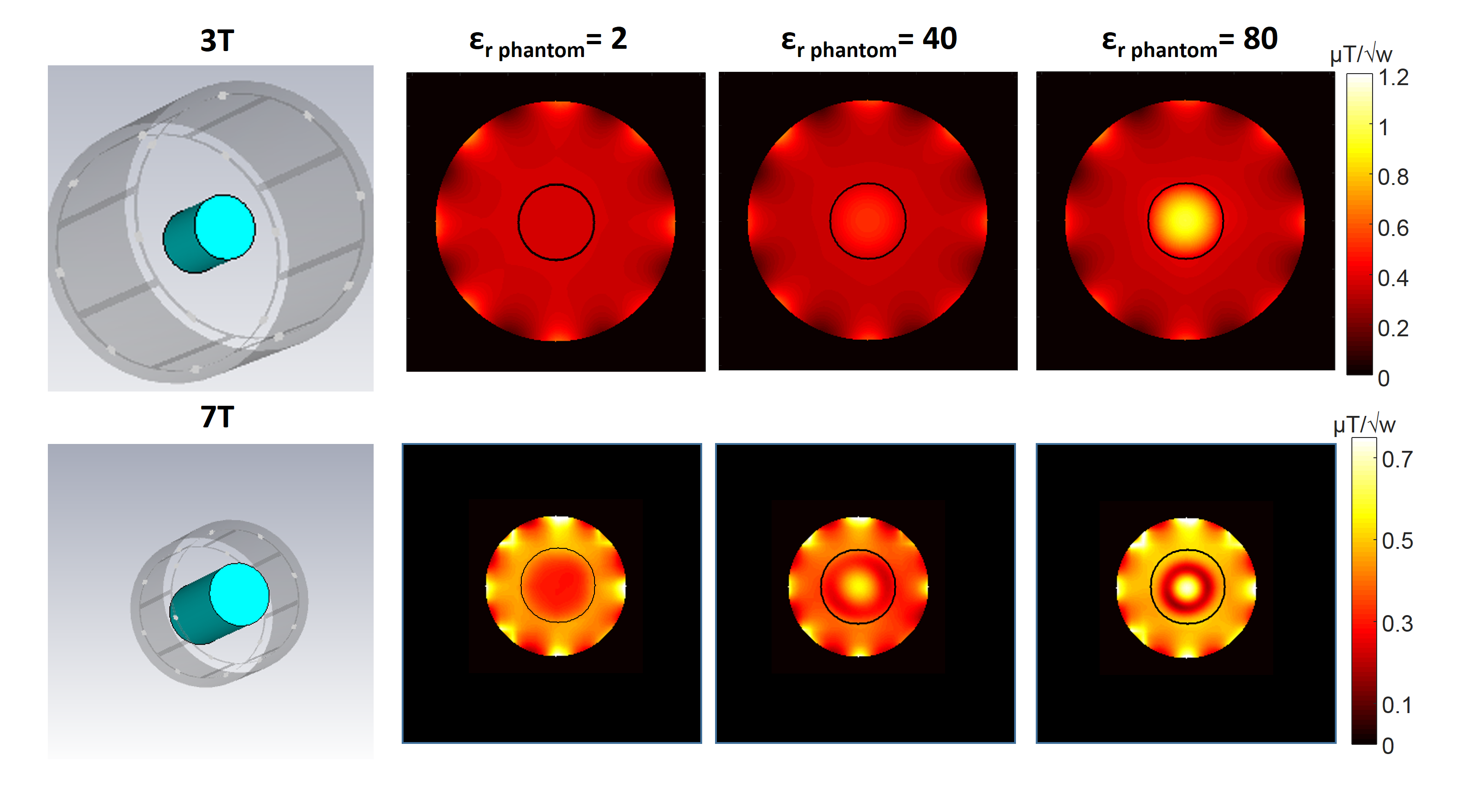
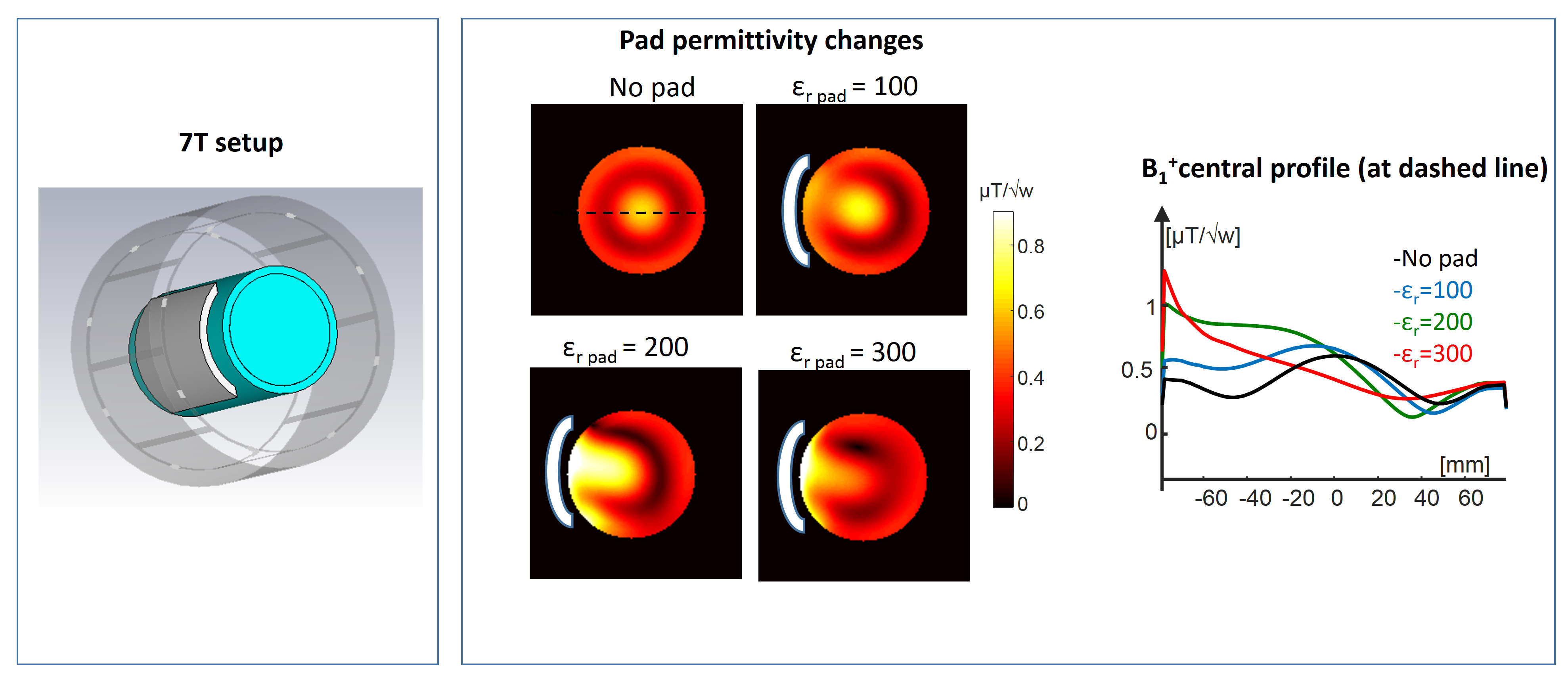
A dielectric material is characterized by its electrical permittivity (also called dielectric constant), which describes the generated electrical polarization in the presence of an electric field and is responsible for storing RF energy1-2 (related to RF transmit efficiency - see Figure 1). Most of the body tissue is made up of dielectric materials with relative to vacuum permittivity (εr) which is frequency dependent2-3. At 128MHz the εr is in the range of 1-100 (bone ~14, fat ~12, muscle ~63, brain gray matter ~73, and CSF ~84)3. As the magnetic field increases the RF wavelength inside the body decreases. Once the wavelength is of the order of the object/region of interest, local interferences appear, also called wavelength effects4-5. These interferences generate local shading and an inhomogeneous RF distribution, which can be observed at 3 T when imaging the abdomen region6 and at 7 T in almost any part of the body7. This is the motivation to study and understand the effects of dielectric materials. Another motivation is the potential use of the dielectric materials for tailoring the RF field distribution. Such RF tailoring is useful in eliminating undesired shading8-9, better SAR management10 and enhancing local efficiency and sensitivity11-12. Dielectric materials are already in common use as pads attached to the region of interest, for example for brain imaging in the Human Connectome Project, when scanning at 7 T13. Dielectric materials are also designed as resonators14, serving as an alternative to common surface or volume coils. Due to the required dimensions, such implementations are more relevant for MRI scanners at fields ≥7 T15. One of the benefits is the simple implementation of such resonators. Better understanding of the effects of dielectric materials can provide new insights in controlling the RF field in MRI.Methods
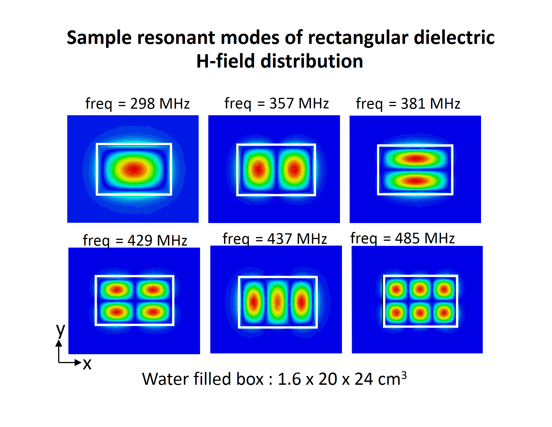
Full estimation of the RF field distribution requires solving the Maxwell set of equations, commonly using dedicated software (simulations shown here were performed using CST Microwave Studio, Darmstadt, Germany). The design of the dielectric material dimensions and properties is mainly based on a forward solution16-17 (calculating the RF field - B1+- for a specific setup), which is repeated to optimize parameters (see for example Figure 2). Published studies include setups of pads made of water10, CaTiO3, and/or BaTiO3 suspensions18-20– having a relative permittivity in the range of 80-300. Mixing different fractions of CaTiO3 or BaTiO3 powder with water will result in different relative permativity εr. A slurry of BaTiO3 beads21 has also been used to reach a higher value of εr (εr = 515). Pad preparation with deionized water is practical in eliminating the signal from the pad itself. Several studies were conducted to analyze the required mixing ratios for generating suspensions with any target permittivity22-23. Recent works also show applications using ultra-high permittivity (500-1000), based on PZT ceramic material24-26. Dielectric materials also support resonant electromagnetic (EM) modes, which depend on the specific shape of the dielectric (see for example Figure 3)27. These are in common use in RF engineering, with a growing interest in MRI. For example, a hollow cylinder was demonstrated to serve as a volumetric resonator15, with implementations at 1H imaging of the calf and wrist and 31P spectroscopy of the calf28. Thin dielectrics can act as resonators similar to surface coil - such resonators were implemented at both 7T and 10.5T29-31.Results
Several groups have demonstrated applications of dielectric materials for improved transmit efficiency, local SNR enhancement, and SAR management. A range of studies was performed with dielectric pads as part of the setup for functional and diffusion MRI at 7T 8,12,32. Studies using dielectric pads at 3 T include abdomen9, cardiac33, and other applications, resulting in better RF distribution. Several works also used dielectric materials as part of the coil assembly, improving the coil efficiency34-36. While most of the work has been done for proton images, feasibility studies included other nuclei as well (13C, 23Na, and 31P). Recent work has shown an enhancement using ultra high permittivity blocks in 1H imaging at 1.5 and 3T24 and in 31P brain spectroscopy at 7T23. Dielectric and cavity resonators using just water show promising results at 7T MRI implemented for limited regions of interest, including: calf, knee, wrist, foot, fingers, and neck 15,37-39. In addition, a multi-transmit resonator was implemented using PZT blocks at 7 T 30,40. A BaTiO3 suspension was also used to show feasibility of a 31P volume resonator at 7 T28.Discussion
Although a range of studies has shown useful applications for dielectric materials, many open questions and challenges remain. Realization of an arbitrary target RF magnetic or electric field is still a challenge. Designs of pads with more complicated geometric shapes are limited, since no simple inverse solution exist. Such studies include pad design with a central hole22 and a work that examines empirically more complex shape for local signal improvement40. Several studies show implementations of dielectric resonators and cavity resonators15,37-39. One of the challenges of such resonators is their detuning, i.e., how to turn off the resonator during receive. One such implementation was shown using a structure of conducting strips and PIN diodes41. Using ceramic blocks (for example uHDC) as dielectric materials has an important advantage of having low conductivity/losses. However, shaping such blocks and achieving flexible setups is challenging. One “workaround” is to connect the blocks using conducting strips. Such connections can generate effectively larger blocks, which can have significant effect on the enhancement42,43. Artificial structures combining dielectric material and conducting material can be the next step in development44,45. Such combinations can achieve structures that are more compact and allow new prospects for RF field control.Acknowledgements
No acknowledgement found.References
1. Jin, Jianming. Electromagnetic analysis and design in magnetic resonance imaging. Vol. 1. CRC press, 1998.
2. Webb, A. G. (2011). Dielectric materials in magnetic resonance. Concepts in magnetic resonance part A, 38(4), 148-184.
3. Gabriel, S., Lau, R. W., & Gabriel, C. (1996). The dielectric properties of biological tissues: II. Measurements in the frequency range 10 Hz to 20 GHz. Physics in medicine & biology, 41(11), 2251.
4. Collins, C. M., Liu, W., Schreiber, W., Yang, Q. X., & Smith, M. B. (2005). Central brightening due to constructive interference with, without, and despite dielectric resonance. Journal of Magnetic Resonance Imaging, 21(2), 192-196.
5. Collins, C. M. (2006). Radiofrequency field calculations for high field MRI. In Ultra high field magnetic resonance imaging(pp. 209-248). Springer, Boston, MA.
6. Chang, K. J., Kamel, I. R., Macura, K. J., & Bluemke, D. A. (2008). 3.0-T MR imaging of the abdomen: comparison with 1.5 T. Radiographics, 28(7), 1983-1998.
7. Vaughan, J. T., Garwood, M., Collins, C. M., Liu, W., DelaBarre, L., Adriany, G., ... & Ugurbil, K. (2001). 7T vs. 4T: RF power, homogeneity, and signal‐to‐noise comparison in head images. Magnetic resonance in medicine, 46(1), 24-30.
8. Yang, Q. X., Mao, W., Wang, J., Smith, M. B., Lei, H., Zhang, X., ... & Chen, W. (2006). Manipulation of image intensity distribution at 7.0 T: passive RF shimming and focusing with dielectric materials. Journal of magnetic resonance imaging, 24(1), 197-202.
9. De Heer, P., Brink, W. M., Kooij, B. J., & Webb, A. G. (2012). Increasing signal homogeneity and image quality in abdominal imaging at 3 T with very high permittivity materials. Magnetic resonance in medicine, 68(4), 1317-1324.
10. Yang, Q. X., Wang, J., Wang, J., Collins, C. M., Wang, C., & Smith, M. B. (2011). Reducing SAR and enhancing cerebral signal‐to‐noise ratio with high permittivity padding at 3 T. Magnetic resonance in medicine, 65(2), 358-362.
11. Brink, W. M., van der Jagt, A. M., Versluis, M. J., Verbist, B. M., & Webb, A. G. (2014). High permittivity dielectric pads improve high spatial resolution magnetic resonance imaging of the inner ear at 7 T. Investigative radiology, 49(5), 271-277.
12. Vaidya, M. V., Lazar, M., Deniz, C. M., Haemer, G. G., Chen, G., Bruno, M., ... & Collins, C. M. Improved detection of fMRI activation in the cerebellum at 7T with dielectric pads extending the imaging region of a commercial head coil. Journal of Magnetic Resonance Imaging.
13. Vu, A. T., Auerbach, E., Lenglet, C., Moeller, S., Sotiropoulos, S. N., Jbabdi, S., ... & Ugurbil, K. (2015). High resolution whole brain diffusion imaging at 7 T for the Human Connectome Project. Neuroimage, 122, 318-331.
14. Kajfez, D., & Guillon, P. (1986). Dielectric resonators. Norwood, MA, Artech House, Inc., 1986, 547 p. No individual items are abstracted in this volume.
15. Aussenhofer, S. A., & Webb, A. G. (2012). Design and evaluation of a detunable water‐based quadrature HEM11 mode dielectric resonator as a new type of volume coil for high field MRI. Magnetic resonance in medicine, 68(4), 1325-1331.
16. Brink, W. M., Remis, R. F., & Webb, A. G. (2016). A theoretical approach based on electromagnetic scattering for analysing dielectric shimming in high‐field MRI. Magnetic resonance in medicine, 75(5), 2185-2194.
17. Van Gemert, J., Brink, W., Webb, A., & Remis, R. (2017). An efficient methodology for the analysis of dielectric shimming materials in magnetic resonance imaging. IEEE transactions on medical imaging, 36(2), 666-673.
18. O'brien, K. R., Magill, A. W., Delacoste, J., Marques, J. P., Kober, T., Fautz, H. P., ... & Krueger, G. (2014). Dielectric pads and low‐B1+ adiabatic pulses: Complementary techniques to optimize structural T1w whole‐brain MP2RAGE scans at 7 tesla. Journal of Magnetic Resonance Imaging, 40(4), 804-812.
19. Teeuwisse, W. M., Brink, W. M., Haines, K. N., & Webb, A. G. (2012). Simulations of high permittivity materials for 7 T neuroimaging and evaluation of a new barium titanate‐based dielectric. Magnetic resonance in medicine, 67(4), 912-918.
20. Snaar, J. E. M., Teeuwisse, W. M., Versluis, M. J., van Buchem, M. A., Kan, H. E., Smith, N. B., & Webb, A. G. (2011). Improvements in high‐field localized MRS of the medial temporal lobe in humans using new deformable high‐dielectric materials. NMR in Biomedicine, 24(7), 873-879.
21. Luo, W., Lanagan, M. T., Sica, C. T., Ryu, Y., Oh, S., Ketterman, M., ... & Collins, C. M. (2013). Permittivity and performance of dielectric pads with sintered ceramic beads in MRI: early experiments and simulations at 3 T. Magnetic resonance in medicine, 70(1), 269-275.
22. O’Reilly, T. P. A., Webb, A. G., & Brink, W. M. (2016). Practical improvements in the design of high permittivity pads for dielectric shimming in neuroimaging at 7 T. Journal of Magnetic Resonance, 270, 108-114.
23. Neves, A. L., Leroi, L., Raolison, Z., Cochinaire, N., Letertre, T., Abdeddaim, R., ... & Malléjac, N. (2018). Compressed perovskite aqueous mixtures near their phase transitions show very high permittivities: New prospects for high‐field MRI dielectric shimming. Magnetic resonance in medicine, 79(3), 1753-1765.
24. Lee, B. Y., Zhu, X. H., Rupprecht, S., Lanagan, M. T., Yang, Q. X., & Chen, W. (2017). Large improvement of RF transmission efficiency and reception sensitivity for human in vivo 31P MRS imaging using ultrahigh dielectric constant materials at 7 T. Magnetic resonance imaging, 42, 158-163.
25. Rupprecht, S., Lee, B. Y., Zhu, X. H., Chen, W., & Yang, Q. X. (2014). Signal-to-Noise Ratio Improvement for MR Proton Spectroscopy at 3T Using a Ultra-high Dielectric Constant (uHDC) Material Sleeve. Proceedings of the International Society for Magnetic Resonance in Medicine, Milan, Italy, 0403.
26. Vaidya, M. V., Sodickson, D. K., Collins, C. M., & Lattanzi, R. (2014, May). Extending the Sensitivity of a Transmit/Receive Radiofrequency Coil with Dielectric Materials at 7 T. In Proc. Intl. Soc. Mag. Reson. Med (Vol. 22, p. 0406).
27. Webb, A. G. (2012). Visualization and characterization of pure and coupled modes in water-based dielectric resonators on a human 7T scanner. Journal of Magnetic Resonance, 216, 107-113.
28. Schmidt, R., & Webb, A. (2016). Characterization of an HEM-Mode Dielectric Resonator for 7-T human phosphorous magnetic resonance imaging. IEEE Transactions on Biomedical Engineering, 63(11), 2390-2395.
29. Aussenhofer, S. A., & Webb, A. G. (2014). An eight-channel transmit/receive array of TE01 mode high permittivity ceramic resonators for human imaging at 7 T. Journal of Magnetic Resonance, 243, 122-129.
30. O'Reilly, T., Ruytenberg, T., & Webb, A. G. (2018). Modular transmit/receive arrays using very‐high permittivity dielectric resonator antennas. Magnetic resonance in medicine, 79(3), 1781-1788.
31. Lagore R. L., DelaBarre L., Yang Q. X., Lanagan M., Eryaman Y., Rupprecht S., Luo W., Lee B.-Y., Zhu X.-H. , Ugurbil K. , Chen W., and Adriany G. High dielectric constant (HDC) disk dipoles for 10.5T imaging. Proc. Intl. Soc. Mag. Reson. Med. 25 (2017), 1128.
32. Schaller, B., Xin, L., O'brien, K., Magill, A. W., & Gruetter, R. (2014). Are glutamate and lactate increases ubiquitous to physiological activation? A 1H functional MR spectroscopy study during motor activation in human brain at 7 Tesla. Neuroimage, 93, 138-145.
33. Brink, W. M., van den Brink, J. S., & Webb, A. G. (2015). The effect of high-permittivity pads on specific absorption rate in radiofrequency-shimmed dual-transmit cardiovascular magnetic resonance at 3T. Journal of Cardiovascular Magnetic Resonance, 17(1), 82.
34. Rupprecht, S., Sica, C. T., Chen, W., Lanagan, M. T., & Yang, Q. X. (2018). Improvements of transmit efficiency and receive sensitivity with ultrahigh dielectric constant (uHDC) ceramics at 1.5 T and 3 T. Magnetic resonance in medicine, 79(5), 2842-2851.
35. Kordzadeh, A., & De Zanche, N. (2018). Optimal-permittivity Dielectric Liners for a 4.7 T Transceiver Array. Magnetic resonance imaging, 48, 89-95.
36. Vaidya, M. V., Deniz, C. M., Collins, C. M., Sodickson, D. K., & Lattanzi, R. (2017). Manipulating transmit and receive sensitivities of radiofrequency surface coils using shielded and unshielded high-permittivity materials. Magnetic Resonance Materials in Physics, Biology and Medicine, 1-12.
37. Bluemink, J. J., Raaijmakers, A. J., Koning, W., Andreychenko, A., Rivera, D. S., Luijten, P. R., ... & van den Berg, C. A. (2016). Dielectric waveguides for ultrahigh field magnetic resonance imaging. Magnetic resonance in medicine, 76(4), 1314-1324.
38. Koning, W., Bluemink, J. J., Langenhuizen, E. A. J., Raaijmakers, A. J., Andreychenko, A., den Berg, C. A. T., ... & Klomp, D. W. (2013). High‐resolution MRI of the carotid arteries using a leaky waveguide transmitter and a high‐density receive array at 7 T. Magnetic resonance in medicine, 69(4), 1186-1193.
39. Aussenhofer, S. A., & Webb, A. G. (2013). High‐permittivity solid ceramic resonators for high‐field human MRI. NMR in Biomedicine, 26(11), 1555-1561.
40. Schmidt, R., & Webb, A. (2016). Improvements in RF Shimming in High Field MRI Using High Permittivity Materials With Low Order Pre-Fractal Geometries. IEEE transactions on medical imaging, 35(8), 1837-1844.
41. Ruytenberg, T., & Webb, A. G. (2017). Design of a dielectric resonator receive array at 7 Tesla using detunable ceramic resonators. Journal of Magnetic Resonance, 284, 94-98.
42. Schmidt, R., Teeuwisse, W., & Webb, A. (2017). Quadrature operation of segmented dielectric resonators facilitated with metallic connectors. Magnetic resonance in medicine, 77(6), 2431-2437.
43. Koolstra, K., Börnert, P., Brink, W., & Webb, A. (2018). Improved image quality and reduced power deposition in the spine at 3 T using extremely high permittivity materials. Magnetic resonance in medicine, 79(2), 1192-1199.
44. Slobozhanyuk, A. P., Poddubny, A. N., Raaijmakers, A. J., van den Berg, C. A., Kozachenko, A. V., Dubrovina, I. A., ... & Belov, P. A. (2016). Enhancement of magnetic resonance imaging with metasurfaces. Advanced materials, 28(9), 1832-1838.
45. Schmidt, R., Slobozhanyuk, A., Belov, P., & Webb, A. (2017). Flexible and compact hybrid metasurfaces for enhanced ultra high field in vivo magnetic resonance imaging. Scientific reports, 7(1), 1678.
Figures