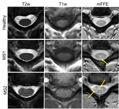
How to Optimise Spinal Cord MR Acquisitions
1Radiology, Vanderbilt University Medical Center, Nashville, TN, United States, 2Vanderbilt Univeristy Institute of Imaging Science, Vanderbilt University Medical Center, Nashville, TN, United States
Synopsis
The goal of this educational presentation is to develop an understanding of where spinal cord MRI is, its optimization, and impediments to future optimization. We will study what spinal cord optimization means and how improvements can have a real, sustained impact on the radiological and scientific community. We will identify needs, both patient and research centered, that could be addressed with improved spinal cord imaging methods. We will close by asking, “if we know what we need to have, and can identify some of the impediments to those needs, how do we optimize a spinal cord examination for maximal impact?”
Background
The spinal cord is a small, largely white matter bundle that exits the brain and is responsible for the majority of community to and from the rest of the body. It has been shown to be clinically eloquent in many diseases, syndromes, and conditions, however, gaining non-invasive information about this anatomical structure has remained difficult. MRI has now been available to the clinical and research community for nearly 50 years, however, the bulk of MRI development still focuses on improving tools and techniques for better, more sensitive/specific assessments of brain structure and function. To the un-initiated, perhaps the spinal cord is merely an extension of the brain, potentially less interesting, or more nefarious, the belief that spinal cord MRI is difficult and a prohibitive anatomy from which to obtain advanced MRI indices.
Conventional wisdom suggests that spinal cord MRI has lagged behind its brain counterpart by approximately 10 years. The tools that exist in the literature in 2018 are tools that have been explored in the brain for many years before (e.g. resting state fMRI, atlases for coregistration/segmentation, MR spectroscopy, etc). However, it is important to note that significant advancement of spinal cord acquisition and analyses tools have occurred and are worth examining further. Additionally, with improved understanding of disease, clinicians often report the need for greater spinal cord tools from which to derive meaningful outcome measures that aid in patient treatment, evaluation of therapies, and assessing overall disease evolution.
The question remains, however, what is needed to optimize a spinal cord examination. Perhaps a better question is, what needs to be done with spinal cord MRI to bring it up to speed with similar studies in the brain, and what are the impediments to doing so. Is the goal to optimize spinal cord imaging to report on similar indices as are seen in the brain, or should we think about a more disruptive paradigm and ask what indices do we need to see in the spinal cord that are clinically relevant and ripe for research interrogation?
Finally, while the desire is often developing acquisitions that generate quantitative indices reflective of important aspects of spinal cord (or, generically, the nervous system), optimization of acquisition strategies need to be considered first. In this presentation we will focus on building up knowledge of some of the current challenges facing spinal cord MRI, address some of the improvements many groups have made in overcoming these challenges, and lastly open up discussion about what are some of the current remaining needs? Can we, through targeted spinal cord optimization, change the conversation surrounding spinal cord MRI and offer real, impactful clinical tools that enrich our understanding of the role the spinal cord plays in many diseases?
Goals of Presentation
We have four primary goals of this presentation: 1) describe the current imaging tools that exist for spinal cord MRI, 2) discuss the impediments to “getting what we want” with improved, optimized spinal cord MRI, 3) identify targets for optimization, and 4) discuss what optimization of spinal cord MRI looks like in context of clinical improvement and research questions.Highlights
Highlights
· Description of current spinal cord MRI both clinically and in the research arena
· What are the current impediments to advanced, or optimized spinal cord MRI
· What are some of the improvements that have already been made
· What are the tools that we need to develop to make real clinical and research impact
· How does one optimize a spinal cord MRI experiment, or clinical evaluation package
· Discussion of disruptive contrasts and how they play a role in spinal cord MRI optimization
