Diffusion Images for Assessing Glymphatic System
1Nagoya University, Japan
Synopsis
Recently introduced “glymphatic system” is a coined word that combines “gl” for glia and “lymphatic” system. In this hypothesis, the perivascular space functions as a conduit for flowing cerebrospinal fluid into brain parenchyma. Activity of the glymphatic system may be evaluated with diffusion images. Lower diffusivity along the perivascular space seems to reflect impairment of the glymphatic system. Diffusion method may be feasible for evaluating the activity of the glymphatic system.
I. Glymphatic system
The lymphatic system is involved in protein waste removal from the body. On the other hand, no lymphatic drainage like the body was present in the brain. Nedergaard and Iliff et al. hypothesized that the perivascular space constitutes a system corresponding to the lymphatic system in the brain and named this the “glymphatic system”, which is a coined word that combines “g” for glia cell and “lymphatic” system. (1, 2).The outline of their hypothesis is as follows. The perivascular space functions as a conduit for cerebrospinal fluid to flow into the brain parenchyma. The driving force of this conduit is arterial pulsation. Cerebrospinal fluid is led to the perivascular space around the artery and enters the interstitial space of the brain tissue through water channels controlled by aquaporin 4 (AQP4), which is distributed in the foot processes of astrocytes that make up the outer wall of the perivascular space. Cerebrospinal fluid entering the interstitial space washes out waste proteins, such as amyloid β, from the tissue. The cerebrospinal fluid that has washed out from between the cells in this way flows into the perivascular space around the veins and is discharged outside the brain. Above mentioned glymphatic system has a characteristic feature, that the function of activity is modified by sleep. This feature was confirmed by an experience on the drainage of amyloid β, which is a protein associated with Alzheimer's disease, causes aggregates interstitially to form amyloid plaques and contributes to disease progression. A report on the excretion of amyloidβ in healthy mice and AQP4 knockout mice showed that natural sleep or anesthesia are associated with a 60% increase in the interstitial space, resulting in a striking increase in exchange of cerebrospinal fluid with interstitial fluid (3).II. Evaluation of glymphatic system: Tracer study
Observation of the glymphatic system has been made with tracer studies in animal experiments (1, 4, 5). Iliff et al. showed that CSF rapidly enters the brain along the cortical pial arteries following labeling of CSF with fluorescent tracers injected into the CSF of the cisterna magna. The dynamics of the glymphatic system were characterized for the first time in vivo using two-photon microscopy in mice (1). In addition to fluorescence materials, intrathecal administration of GBCM was also used as a tracer in later animal studies. An experimental rat study with whole-brain imaging following intrathecal GBCM administration allowed identification of two key influx nodes at the pituitary and pineal gland recesses (4). However, intrathecal administration of tracers in humans has not been performed except for several well-designed studies (6) and an accident report (7). Naganawa et al. observed enhancement of the perivascular space and CSF space on heavily T2-weighted FLAIR images 4 hours after intravenous injection of GBCM (8); however, no method for tracing intravenously administered GBCM with MRI has been established. In addition, even if a tracer method to evaluate the glymphatic system in living humans is established, tracer studies of the glymphatic system require hours to follow the distribution of the tracer within the brain, and monitoring the activity of the glymphatic system in real time is not possible.III. Evaluation of glymphatic system: Non-tracer study by diffusion images.
There are several trial to evaluate the activity of the glymphatic system using diffusion image on MRI. One of the trial is a method called “diffusion tensor image analysis along the perivascular space (DTI-ALPS)” for evaluating the activity of the glymphatic system in human brain by using diffusion images. In this method, the motion of water molecule in the direction of the perivascular space are evaluated by measuring diffusivity using the diffusion tensor dataset(9). At the level of the lateral ventricle body, the medullary veins run perpendicular to the ventricular wall, and the perivascular space runs in the same direction as the medullary veins, which is the right-left direction (x-axis). On the plane of this area, projection fibers run in the head-foot (z-axis) direction, mainly adjacent to the lateral ventricle, and superior longitudinal fascicles (SLFs) representing association fibers in the current study run in the anterior-posterior (y-axis) direction outside the projection fibers. Outside the SLFs, subcortical fibers run mainly in the right-left (y-axis) direction in subcortical areas. Consequently, in this area, the perivascular space runs perpendicular to the projection fibers and SLFs. This conformation of the perivascular space and major fibers in this area allows nearly independent analysis of the diffusivity along the direction of the perivascular space because major fiber tracts do not run parallel to the direction of the perivascular space (Figure 1 a,b). When common changes in the diffusivity in the right-left direction (x-axis) in both the projection fiber area and association fiber area are present, the change is a result of the alteration in the diffusivity in this direction that agrees with the direction of the perivascular space. Evaluation of Alzheimer’s disease patients by using DTI-ALPS method were carried out (Figure 2 a-c). The results indicated significant negative correlation between MMSE scores and the diffusivity along the projection fibers and association fibers. This result can be explained by white matter degeneration in the projection or association fibers due to AD or MCI. In contrast, the study showed a significant positive correlation between the diffusivity along the perivascular space and the MMSE score, indicating impaired water diffusivity in the direction of the perivascular space in relation to AD severity in areas with projection or association fiber dominance. ALPS-score in order to evaluate the activity of the glymphatic system in individual cases was calculated (Figure 2 d). This score is provided by the ratio of two sets of diffusivity value which are perpendicular to dominant fibers in the tissue. It is hypothesized that the ratio of them would express the influence of the water diffusion along perivascular space which will reflect activity of the glymphatic system in the individual cases. There were significant positive correlation between the ALPS-index and the MMSE scores on the b=1000s/mm2 datasets. This result would indicate that the ALPS-index can be used for evaluating the activity of the glymphatic system in the individual cases.Acknowledgements
No acknowledgement found.References
1. Iliff JJ, Wang M, Liao Y, et al. A paravascular pathway facilitates CSF flow through the brain parenchyma and the clearance of interstitial solutes, including amyloid beta. Sci Transl Med. 2012;4(147):147ra11.
2. Nedergaard M, Goldman SA. Brain Drain. Sci Am. 2016;314(3):44-9.
3. Xie L, Kang H, Xu Q, et al. Sleep drives metabolite clearance from the adult brain. Science. 2013;342(6156):373-7.
4. Iliff JJ, Lee H, Yu M, et al. Brain-wide pathway for waste clearance captured by contrast-enhanced MRI. J Clin Invest. 2013;123(3):1299-309.
5. Jessen NA, Munk AS, Lundgaard I, Nedergaard M. The Glymphatic System: A Beginner's Guide. Neurochem Res. 2015;40(12):2583-99.
6. Oner AY, Barutcu B, Aykol S, Tali ET. Intrathecal Contrast-Enhanced Magnetic Resonance Imaging-Related Brain Signal Changes: Residual Gadolinium Deposition? Invest Radiol. 2017;52(4):195-7.
7. Samardzic D, Thamburaj K. Magnetic resonance characteristics and susceptibility weighted imaging of the brain in gadolinium encephalopathy. J Neuroimaging. 2015;25(1):136-9.
8. Naganawa S, Nakane T, Kawai H, Taoka T. Gd-based Contrast Enhancement of the Perivascular Spaces in the Basal Ganglia. Magn Reson Med Sci. 2016;16(1):61-5.
9. Taoka T, Masutani Y, Kawai H, et al. Evaluation of glymphatic system activity with the diffusion MR technique: diffusion tensor image analysis along the perivascular space (DTI-ALPS) in Alzheimer's disease cases. Jpn J Radiol. 2017;35(4):172-8.
Figures
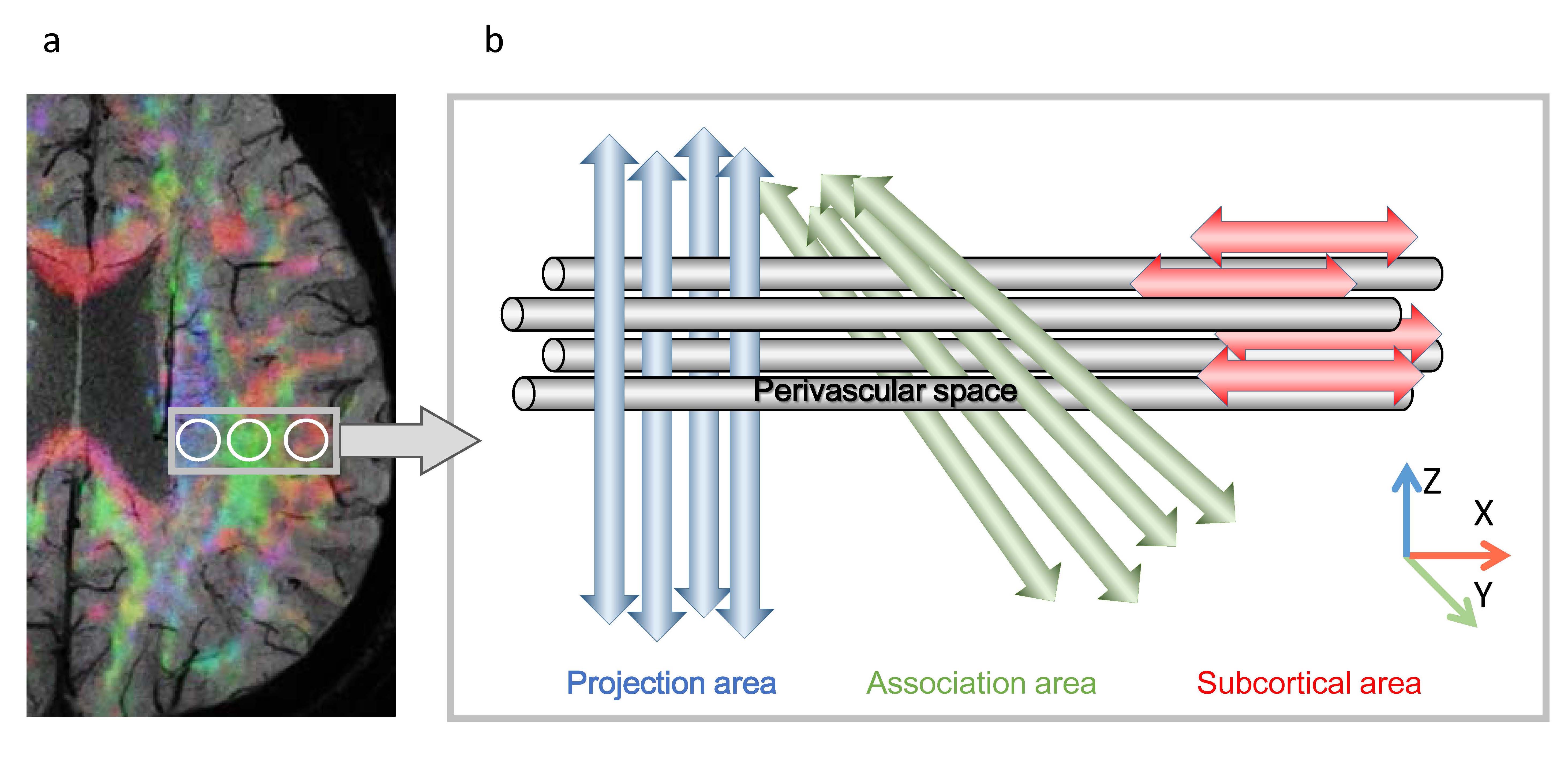
Figure 1: Concept for DTI-ALPS method
a.Superimposed color display of DTI on SWI indicating the distribution of projection, association and the subcortical area.
b.Schematic indicating the relationship between the direction of the perivascular space (gray cylinder) and the directions of the fibers.
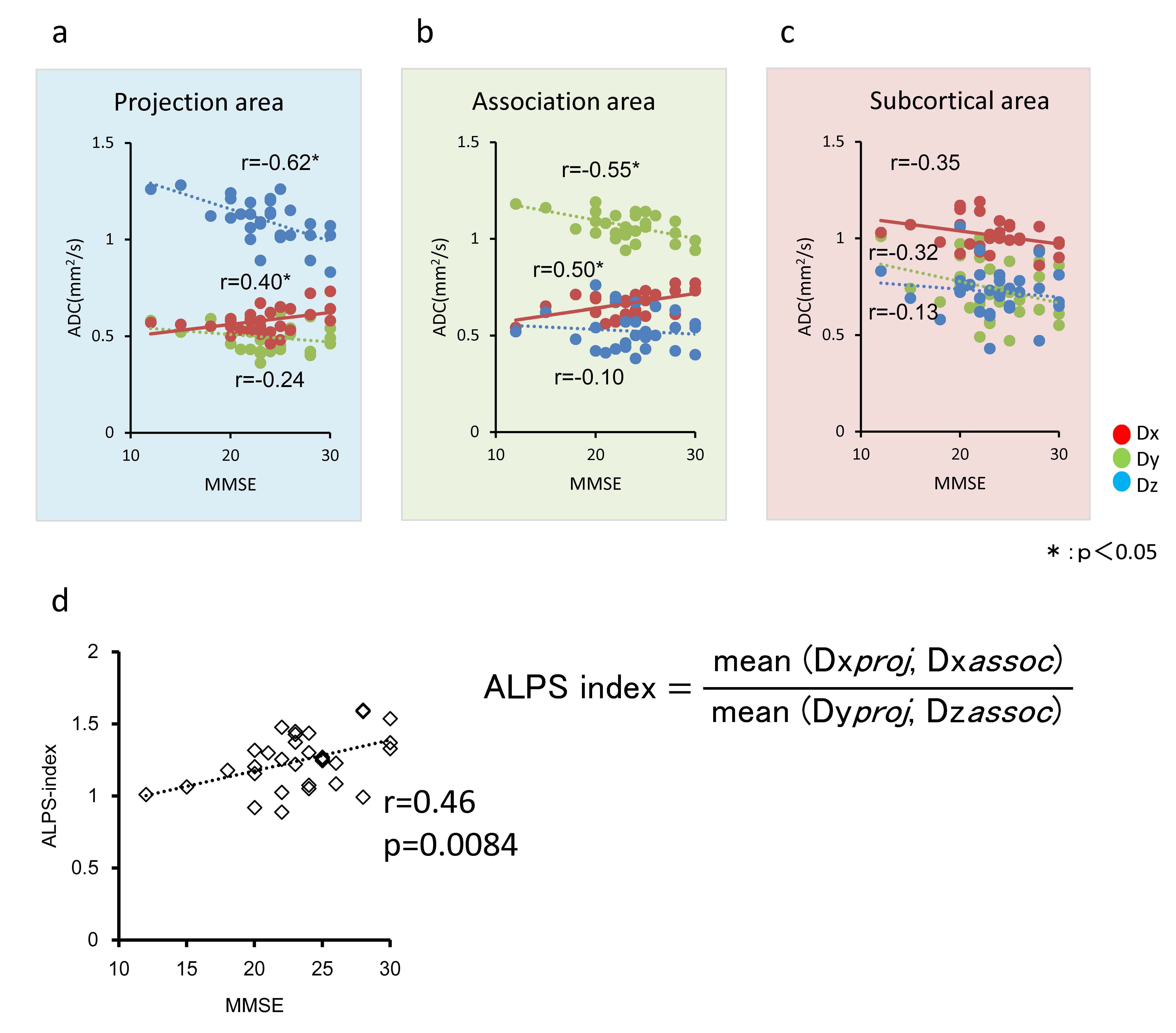
Figure 2: Correlation between directional diffusivity and MMSE scores
a: projection area, b: association area, c: subcortical area
Significant positive correlation between the diffusivity along the perivascular space (x-axis) and the MMSE scores.
d: Correlation between ALPS-index and MMSE.
There were significant positive correlation between the ALPS-index and MMSE scores