Sustainable Low-field MRI for Point of Care Diagnostics
1Center for Neural Engineering, Penn State University, University Park, PA, United States
Synopsis
Low-field MRI has been technically feasible for decades, but clinically diagnostic MRI has been dominated by high-field technology. Thus the vast majority of people on earth have no foreseeable access to MRI. We are developing diagnostic low-field MRI, by addressing the hardware and algorithmic challenges that have been barriers to clinical use. An overarching principle is that our designs can be assembled, operated and maintained in developing countries. We initially focus on hydrocephalus – the most common condition in children worldwide that requires brain imaging and neurosurgical treatment. We will define sustainability for such diagnostics using an integrated optimization approach.
Of the estimated 400,000 new cases of hydrocephalus each year, the majority are in the developing world. There are 3 features of infant hydrocephalus that render it ideal for low-field low-resolution MRI: 1) water is the strongest signal for proton-based MRI, 2) we need to segment brain and fluid for diagnosis and treatment planning, but tissue contrast and high-resolution is unnecessary, and 3) although we need to image adult head sized fields of view, the small infant body simplifies design challenges.
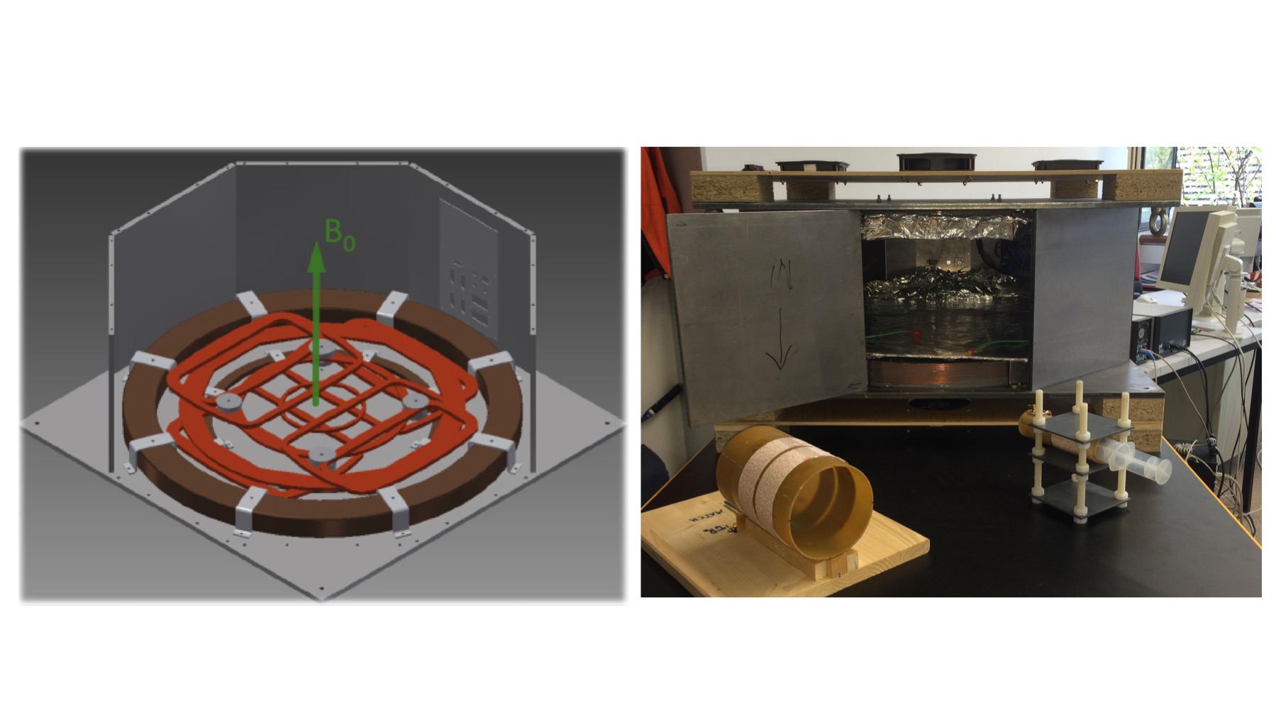
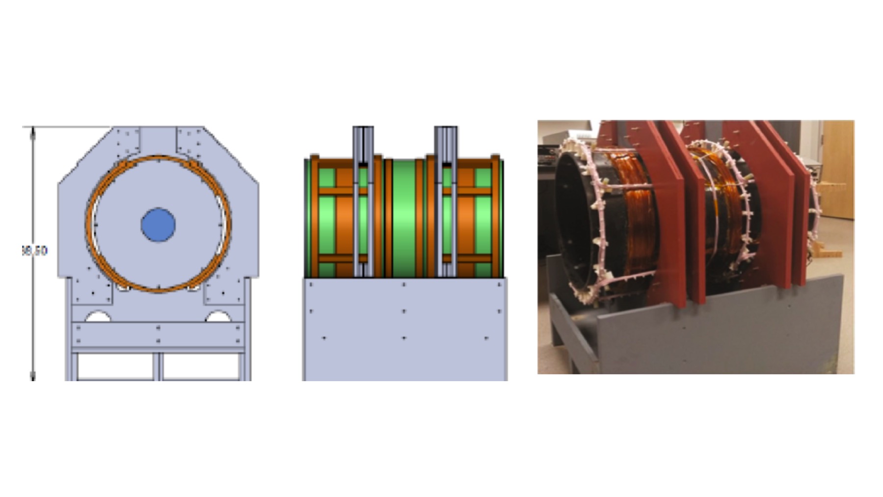
We are proceeding along 3 separate low-cost cryogen-free hardware directions: 1) constant Bo field electromagnet design with steel shields that boost the strength of the Bo (Lother et al 2016, Figure 1), 2) a pre-polarization electromagnet design (Obungoloch et al 2018, Figure 2), and 3) Halbach array designs (Cooley et al 2018). Our initial implementation in Africa is based on the prepolarization design, scaled up in size to admit the adult sized head of the infant with hydrocephalus, but accommodating the infant sized body and shoulders to simplify design requirements. Our intention is to transition to designs that avoid some of the constraints of prepolarization as alternative device designs become available for implementation.
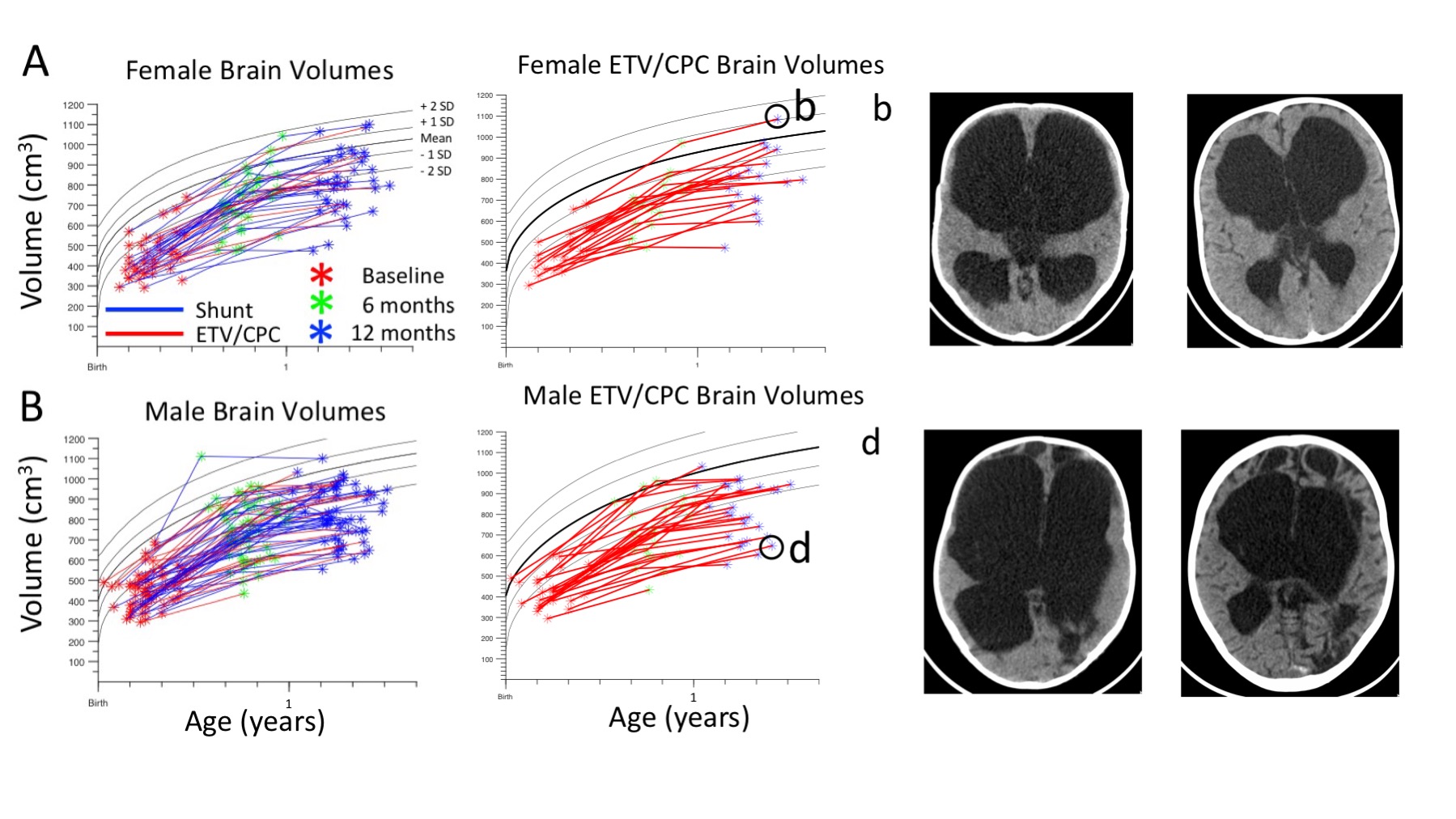
Because such devices should operate in the absence of radiologists, we are developing automated machine learning algorithms for pre- and post-operative diagnostics, as well as image enhancement (Cherukuri et al 2017). We are further developing brain volumetrics compatible with low-resolution (Peterson et al 2018). Following the recent completion of the early childhood growth curves, we have demonstrated both grow-arrest and catch-up growth in infant brains related to increased intracranial pressure before, and reduced pressure following, surgical intervention (Kulkarni et al 2017). Such volumetrics to guide hydrocephalus therapy can be obtained at low resolution.
Lastly, we will describe an attempt to define sustainability for such diagnostics using an integrated optimization approach.
Acknowledgements
Supported by the US National Institutes of Health Director’s Pioneer Award 5DP1HD086071, US National Institutes of Child Health and Development grant 2R01HD085853, and The Netherlands Organisation for Scientific Research - Science for Global Development program NWO-WOTRO W 07.303.101.References
Cherukuri, V, Ssenyonga P, Warf BC, Kulkarni AV, Monga V, Schiff SJ. Learning Based Segmentation of CT Brain Images: Application to Post-Operative Hydrocephalic Scans. IEEE Transactions on Biomedical Engineering, 2017 in press. 10.1109/TBME.2017.2783305
Cooley, C. Z., Haskell, M. W., Cauley, S. F., Sappo, C., Lapierre, C. D., Ha, C. G., Stockmann, J. P., and Wald, L. L. (2018). Design of Sparse Halbach Magnet Arrays for Portable MRI Using a Genetic Algorithm. IEEE Transactions on Magnetics, 54(1), 1-12. doi: 10.1109/tmag.2017.2751001
Kulkarni AV, Schiff SJ, Mbabazi-Kabachelor E, Mugamba J, Ssenyonga P, Donnelly R, Levenbach J, Monga V, Peterson M, MacDonald M, Cherukuri V, Warf BC. Endoscopic Treatment versus Shunting for Infant Hydrocephalus in Uganda, New England Journal of Medicine, 377:2456-64, 2017. doi: 10.1056/NEJMoa1707568. PMID: 29262276
Lother S, Schiff SJ, Neuberger T, Jakoba PM, Fidler F, Design of a mobile, homogeneous and efficient electromagnet with a large field-of-view for neonatal low field MRI, Magnetic Resonance Materials in Physics, Biology and Medicine, (2016) 29:691–698. DOI 10.1007/s10334-016-0525-8. PMID: 26861046
Obungoloch, J., Harper, J. R., Consevage, S., Savukov, I. M., Neuberger, T., Tadigadapa, S., and Schiff, S. J. (2018). Design of a sustainable prepolarizing magnetic resonance imaging system for infant hydrocephalus. Magnetic Resonance Materials in Physics, Biology and Medicine. doi: 10.1007/s10334-018-0683-y. PMID: 29644479
Peterson M, Warf BC, Schiff SJ. Normative Human Brain Volume Growth. Journal of Neurosurgery Pediatrics, Published online March 2, 2018; DOI: 10.3171/2017.10.PEDS17141. PMID: 29498607
Figures