5667
Preclinical MR spectroscopy for studies of in vivo myocardial energy metabolism in heart failure1Singapore Bioimaging Consortium, Agency for Science, Technology, and Research (A*STAR), Singapore, Singapore, 2Department of Radiology, Academic Medical Center, Amsterdam, Netherlands, 3Biomedical Engineering, Eindhoven University of Technology, Eindhoven, Netherlands, 4Radiology, University Medical Center Utrecht, Utrecht, Netherlands
Synopsis
Changes in myocardial energy metabolism have been thought to contribute to the development of heart failure. Here, we will review methods and applications of cardiac 1H MRS, 31P MRS, and hyperpolarized 13C MRS techniques in probing myocardial energy metabolism in vivo in small animal models of heart failure.
Purpose
To review various magnetic resonance spectroscopy (MRS) techniques for the study of in vivo myocardial energy metabolism in rodent models of heart failure.Outline of content
- Brief overview of myocardial energy metabolism
- Technical considerations for in vivo cardiac MRS in small animals
- 1H MRS to measure myocardial lipids and total creatine pool size
- 31P MRS to measure myocardial energy homeostasis
- Hyperpolarized 13C MRS to measure substrate utilization
Summary
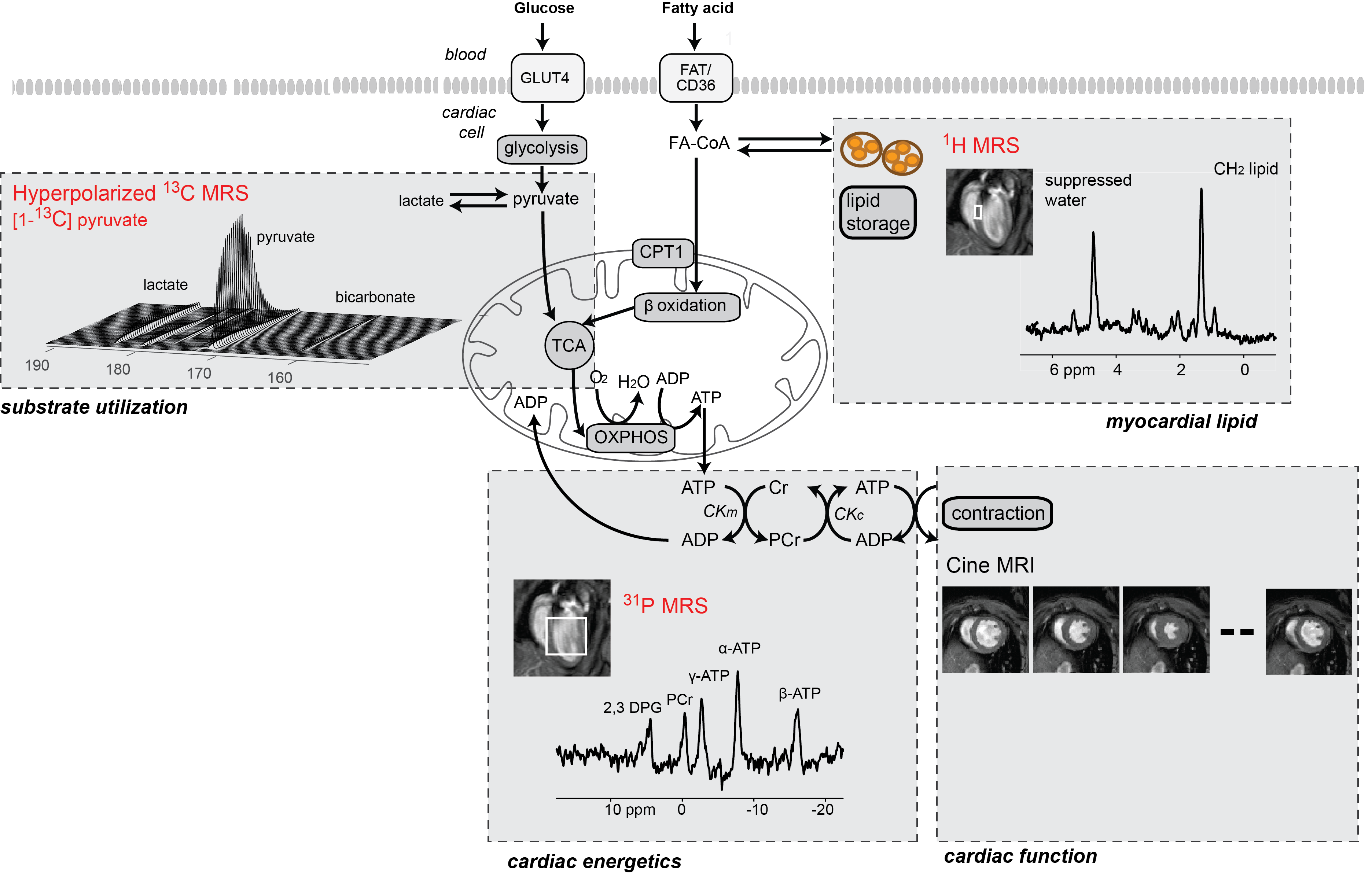
Alterations in myocardial energy metabolism have been implicated in the pathogenesis of heart failure. Studies have shown that the balance of substrate use (i.e. fatty acids, glucose, ketone bodies) is disturbed in heart failure.1-3 While our knowledge of myocardial substrate metabolism largely originates from perfused hearts, the ex vivo setup does not completely mimic the in vivo situation, where substrate availability can change depending on disease conditions. In this case, MRS offers a powerful tool to study myocardial energy metabolism in vivo (Figure 1), which also allows longitudinal studies to map changes during disease progression. Furthermore, cardiac cine imaging can also be implemented to correlate the MRS findings with cardiac function.
In this educational e-poster, we will review various MRS techniques that have been applied to study myocardial energy metabolism during the development of heart failure in small animals by our groups4 and others. We will discuss 1H MRS to measure myocardial lipids and total creatine pool size, 31P MRS to measure cardiac energy homeostasis, and hyperpolarized 13C MRS to study substrate utilization in real time.
Acknowledgements
No acknowledgement found.References
- Glatz, J., et al. Cardiovasc. Drugs Ther. 2006; 20(6):471-476.
- Abdurrachim, D., et al. Cardiovasc. Res. 2017; 113(10):1148-1160.
- Aubert, G., et al. Circulation 2016; 133: 698–705.
- Bakermans, A. J., et al. Prog. Nucl. Magn. Reson. Spectrosc. 2015; 88-89:1-47.
Figures