5656
An Update on Synthetic MRI in Pediatric Brains; Including a Review of MR Quantification Method, Features of the SyMRI Software, and Clinical ApplicationsChristina Andica1, Akifumi Hagiwara1,2, Moeko Horita1,3, Misaki Nakazawa1, Tomoko Maekawa1,2, Saori Koshino1,2, Masaaki Hori1, Lydia Chougar4, and Shigeki Aoki1
1Department of Radiology, Juntendo University Graduate School of Medicine, Tokyo, Japan, 2Department of Radiology, Graduate School of Medicine, The University of Tokyo, Tokyo, Japan, 3Department of Radiological Sciences, Graduate School of Human Health Sciences, Tokyo Metropolitan University, Tokyo, Japan, 4Department of Radiology, Hôpital Cochin, Paris, France
Synopsis
The use of synthetic MRI, a multi-parametric quantitative MRI, in the pediatric population has been increased dramatically in the recent years showing promising results. With this approach, it is possible to synthesize any contrast-weighted images, to obtain automatic brain segmentation and volumetry, and to evaluate the myelin in a single scan. The image quality of synthetic images has been shown comparable to that of conventional MR images, and the features of synthetic MRI might be used to investigate children brain development and diseases. In this exhibit, we aim to show the recent updates of synthetic MRI in pediatric brain imaging.
Purpose
Synthetic MRI with QRAPMASTER (quantification of relaxation times and proton density by multiecho acquisition of a saturation-recovery using turbo spin-echo readout) pulse sequence enables the quantification of R1 and R2 relaxation rates (the inverses of T1 and T2 relaxation times, respectively), proton density (PD), and B1 field in a single scan in about 6 minutes for full head coverage with good accuracy and reproducibility.1, 2 Moreover, reconstruction of various contrast-weighted images,1 automatic brain tissue segmentation and volumetry,3 and measurement of myelin4 can be performed retrospectively using dedicated post-processing SyMRI (SyntheticMR, Linköping, Sweden) software. The use of synthetic MRI may overcome the limitations of conventional MRI in pediatric brain imaging, especially in terms of scanning time, qualitative interpretation, and MR scanner parameters and settings dependence. This exhibit provides the underlying principles and the recent updates of synthetic MRI in pediatric brain imaging.Outline of content
- The basic principles of QRAPMASTER; a multislice, multi-echo, multi-delay acquisition.1, 2
- The features of synthetic MRI:1 (A) Patient-to-patient based tailored contrast-weighted imaging, any contrast-weighted imaging can be synthesized by manipulating acquisition parameters including TR, TE, and TI. (B) Automated brain tissue segmentation and volumetry, white matter (WM), gray matter (GM), cerebrospinal fluid (CSF), non-WM/GM/CSF, brain parenchymal volume, and intracranial volume. (C) Myelin measurement; myelin volume and myelin volume fraction.
- The image quality of synthetic MRI in children brain.5, 6
- Clinical applications of synthetic MRI in the evaluation of brain development7, 8 and pathologies,9-11 for example, Sturge-Weber syndrome, glioma, mild encephalopathy with reversible splenial lesion (MERS), meningitis, multiple sclerosis, tuberous sclerosis, etc.
- The limitations and pitfalls of SyMRI: (A) Lower image quality and partial volume effects of synthetic FLAIR image.6 (B) Sub-optimal WM and GM segmentation in young children.8
Summary
Here, we summarize and review synthetic MRI use in the pediatric brain. With the approach of synthetic MRI, faster, objective, and individualized examination can be achieved in daily clinical practice of pediatric brain imaging.Acknowledgements
The authors have no conflict of interest to disclose.References
- Hagiwara A, Warntjes M, Hori M, et al. SyMRI of the Brain: Rapid Quantification of Relaxation Rates and Proton Density, With Synthetic MRI, Automatic Brain Segmentation, and Myelin Measurement. Invest Radiol 2017
- Warntjes JB, Leinhard OD, West J, et al. Rapid magnetic resonance quantification on the brain: Optimization for clinical usage. Magn Reson Med 2008;60:320-9
- West J, Warntjes JB, Lundberg P. Novel whole brain segmentation and volume estimation using quantitative MRI. Eur Radiol 2012;22:998-1007
- Warntjes M, Engstrom M, Tisell A, et al. Modeling the Presence of Myelin and Edema in the Brain Based on Multi-Parametric Quantitative MRI. Front Neurol 2016;7:16
- Lee SM, Choi YH, Cheon JE, et al. Image quality at synthetic brain magnetic resonance imaging in children. Pediatr Radiol 2017
- West H, Leach JL, Jones BV, et al. Clinical validation of synthetic brain MRI in children: initial experience. Neuroradiology 2017;59:43-50
- Kim HG, Moon WJ, Han J, et al. Quantification of myelin in children using multiparametric quantitative MRI: a pilot study. Neuroradiology 2017
- McAllister A, Leach J, West H, et al. Quantitative Synthetic MRI in Children: Normative Intracranial Tissue Segmentation Values during Development. AJNR Am J Neuroradiol 2017
- Andica C, Hagiwara A, Nakazawa M, et al. The Advantage of Synthetic MRI for the Visualization of Early White Matter Change in an Infant with Sturge-Weber Syndrome. Magn Reson Med Sci 2016;15:347-48
- Hagiwara A, Nakazawa M, Andica C, et al. Dural Enhancement in a Patient with Sturge-Weber Syndrome Revealed by Double Inversion Recovery Contrast Using Synthetic MRI. Magn Reson Med Sci 2016;15:151-2
- Andica C, Hagiwara A, Nakazawa M, et al. Synthetic MR Imaging in the Diagnosis of Bacterial Meningitis. Magn Reson Med Sci 2017;16:91-92
Figures
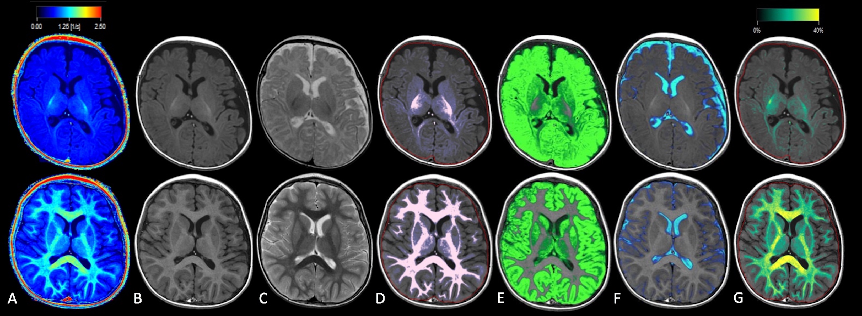
Figure 1. The use of synthetic MRI in the brains of a 3-month-old male infant (first row) and a 2-year-old boy (second row). SyMRI quantitative maps [only a R1 map is shown (A)], synthetic T1WI (B), synthetic T2WI (C), white matter segmentation (D), gray matter segmentation (E), cerebrospinal fluid segmentation (F), and myelin partial volume map (G).