5643
Simultaneous T1 and T2 parametric mapping and synthetic MRI with IR-prepared echo-split gradient-echo and spin-echo imaging and parametric POCSMUSE reconstruction1Department of Biomedical Sciences and Engineering, National Central University, Taoyaun, Taiwan, 2Department of Biomedical Engineering, University of Arizona, Tucson, AZ, United States
Synopsis
Our novel simultaneous T1 and T2 mapping framework, integrating IR-prepared echo-split GRASE acquisition and parametric POCSMUSE reconstruction, has the following advantages. First, T1 and T2 maps can be derived from four sets of single-shot IR-prepared echo-split GRASE data, with very high scan efficiency. Second, T1 and T2 relaxation time constants can be accurately measured by the parametric POCSMUSE algorithm, which models multiplexed signals across CPMG echoes and multiple IR-prepared data of multi-echo-pathway. Third, synthetic multi-contrast images can be generated from the measured parametric maps.
Introduction
Conventional parametric mapping for clinical exams is limited by: 1) long scan time, and 2) inaccurate T2 mapping due to overlapped of multiple echo pathways1. It is, therefore, often impossible to acquire a complete set of high-quality T1 and T2 parametric maps in clinical sessions due to time constraints.
To address these limitations, the newly developed highly-efficient MR imaging technique enables simultaneous T1 and T2 mapping, providing high-quality parametric map with high scan efficient (e.g., < 5 seconds per slice: compatible with multi-slice inversion recovery preparation scheme). The proposed technique can also produce synthetic multi-contrast images, including proton-density-weighted (PDW), T1-weighted (T1W), T2-weighted (T2W) and fluid attenuation inversion recovery (FLAIR) images, from the measured T1 and T2 maps.
Methods
Recently we developed a high-speed T2 parametric mapping framework2 including 1) a single-shot echo-split GRASE (ES-GRASE) imaging sequence for data acquisition with reduced contamination from unwanted echo pathways and 2) parametric POCSMUSE reconstruction measuring an accurate T2 map by incorporating signals from residual echo pathways. We propose to further extend the framework to enable simultaneous T1 and T2 mapping by an IR-prepared ES-GRASE imaging sequence and improved reconstruction.
The proposed framework consists of two modules:
Data Acquisition - IR-prepared ES-GRASE imaging sequence
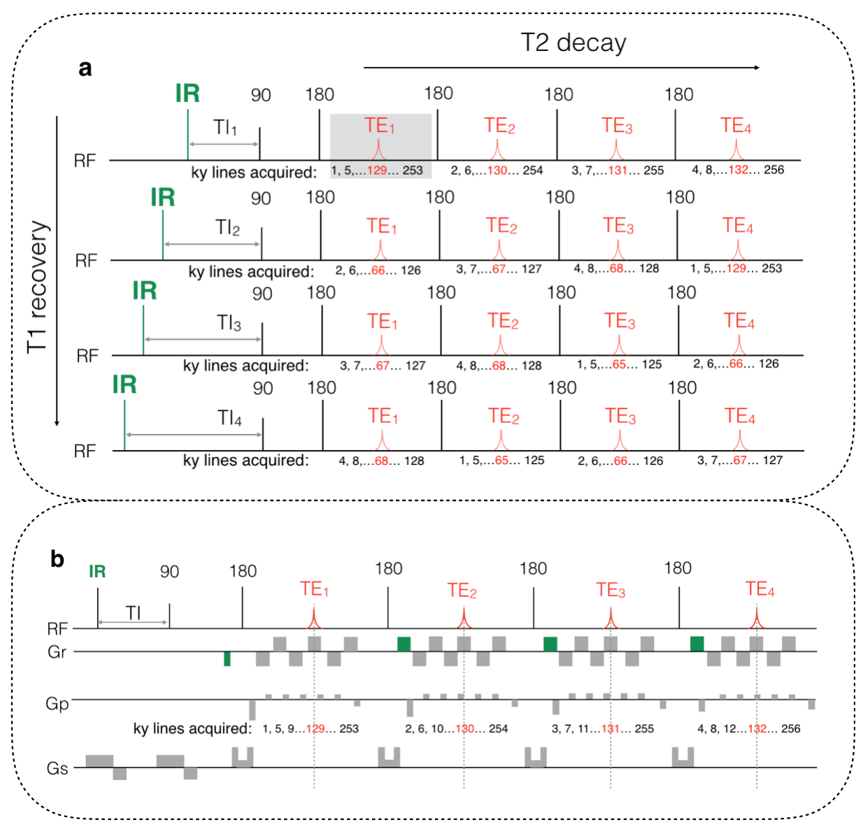
The proposed highly-efficient sequence is illustrated in Figure 1. The sequence includes multiple IR-prepared ES-GRASE acquisitions corresponding to different inversion times (TIs). In each IR-prepared ES-GRASE acquisition (Figure 1b), EPI readout trains in each CPMG echo acquires 1/4 k-space data corresponding to different echo times (TEs). Therefore, the multiple acquisitions (Figure 1a) provide data of 16 echoes with different T2 weightings (across CPMG echoes) and T1 weightings (across multiple IR-prepared datasets) for simultaneous T1 and T2 mapping.
Data Reconstruction - parametric POCSMUSE reconstruction
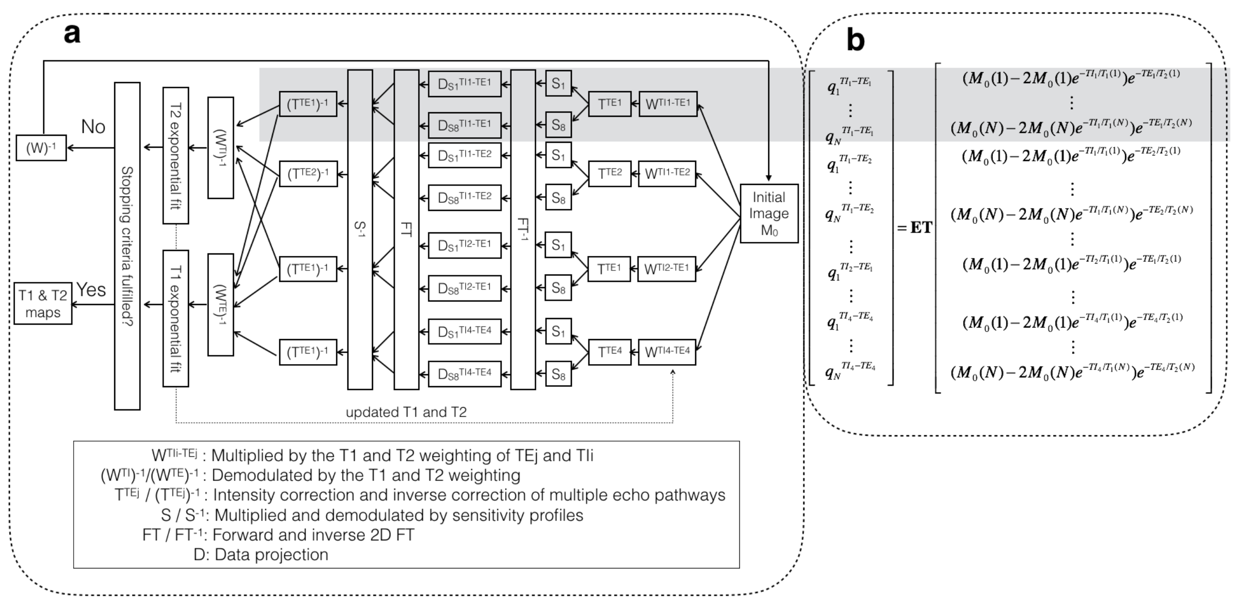
The data from the multiple acquisitions of IR-prepared ES-GRASE sequence can be represented by a multiplexed signal model (see equation in Figure 2b) 2,3, in which $$$Q$$$ is a vector containing acquired k-space signal from 16 echoes at four TEs with four TIs; $$$E$$$ is the encoding matrix with Fourier operators and sensitivity profiles; $$$T$$$ is the matrix containing intensity correction from residual echo pathways; $$$M_0$$$ is a vector containing unknown primary echo signal corresponding to four TEs with four TIs. With the prior knowledge of slice profiles, this non-linear equation can be solved with an iterative parametric POCSMUSE method (see Figure 2a). The POCSMUSE procedures are iterated until a set of a full-FOV T1 and T2 map are produced.
Our novel framework was evaluated with human MRI data obtained from a 3 Tesla scanner. First, four sets of single-shot IR-prepared echo-split GRASE data with four TI = 50, 200, 600, 2000 ms and four CPMG echoes (TE = 75, 145, 215 and 285 ms) were acquired with an 8-channel coil with matrix size = 256×256, FOV = 24x24 cm2, and slice thickness = 8 mm. The data were processed with the parametric POCSMUSE method to produce T1, T2 maps and a PDW image, from which synthetic T1W, T2W, T1-FLAIR and T2-FLAIR images were generated.
Second, four sets of fully-sampled 4-shot spin-echo EPI data were obtained with TE = 75, 145, 215 and 285 ms, respectively. Another four sets of fully-sampled IR-prepared 4-shot spin-echo EPI data with TE = 75 ms and TI = 50, 200, 600, 2000 ms were also acquired respectively. These 4-shot EPI data, containing signals from only the primary spin-echo pathway and with the same geometric distortion as ES-GRASE data, were used as the ground truth in evaluating the developed methods.
Results
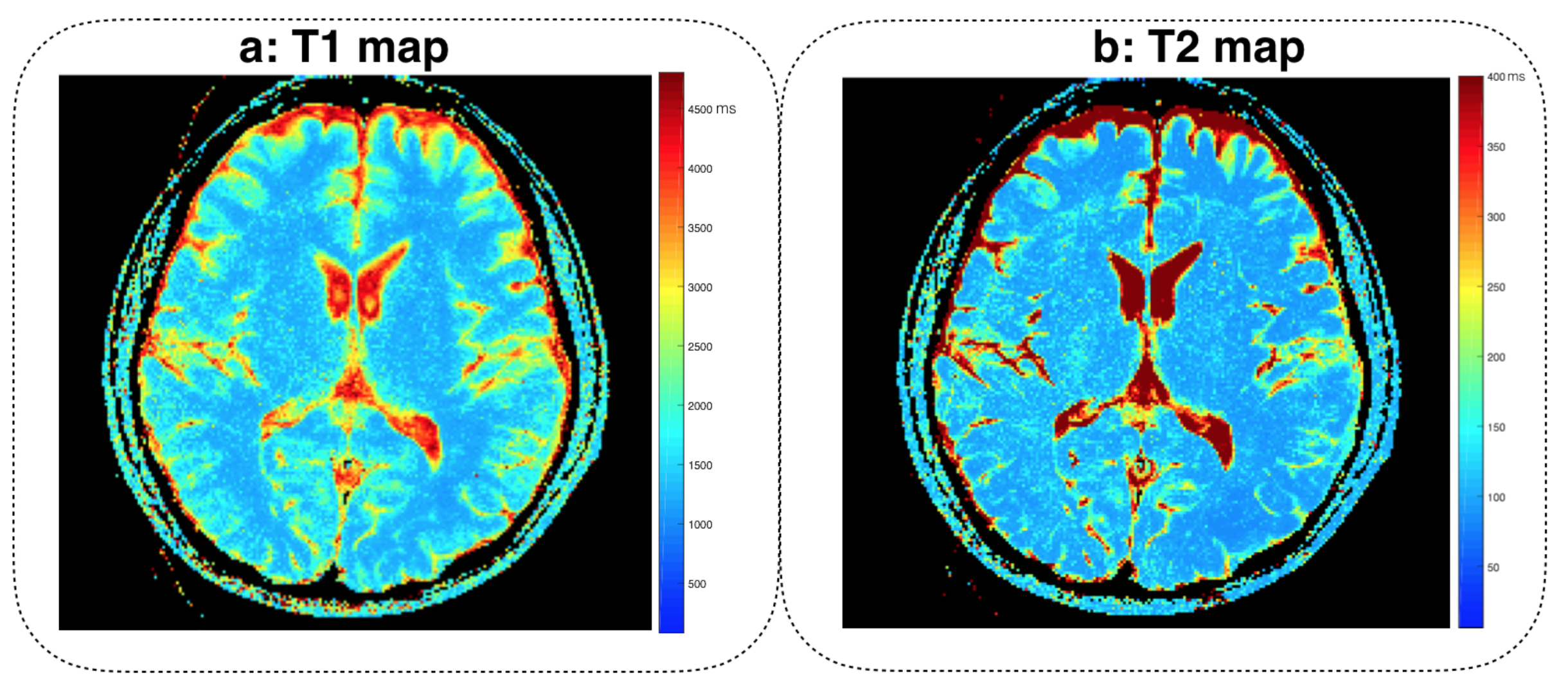
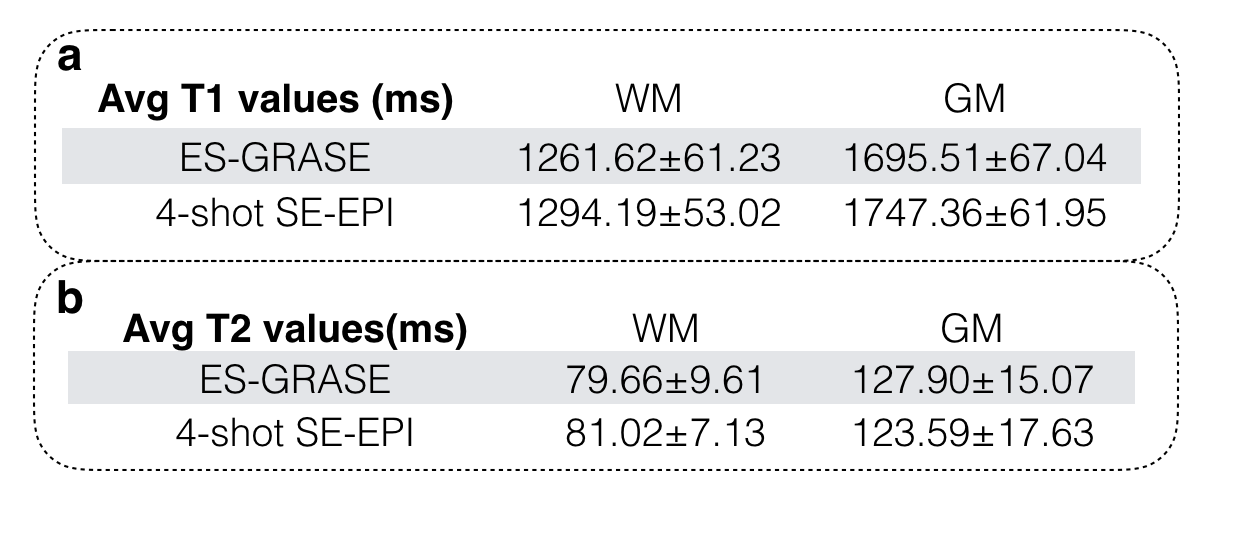
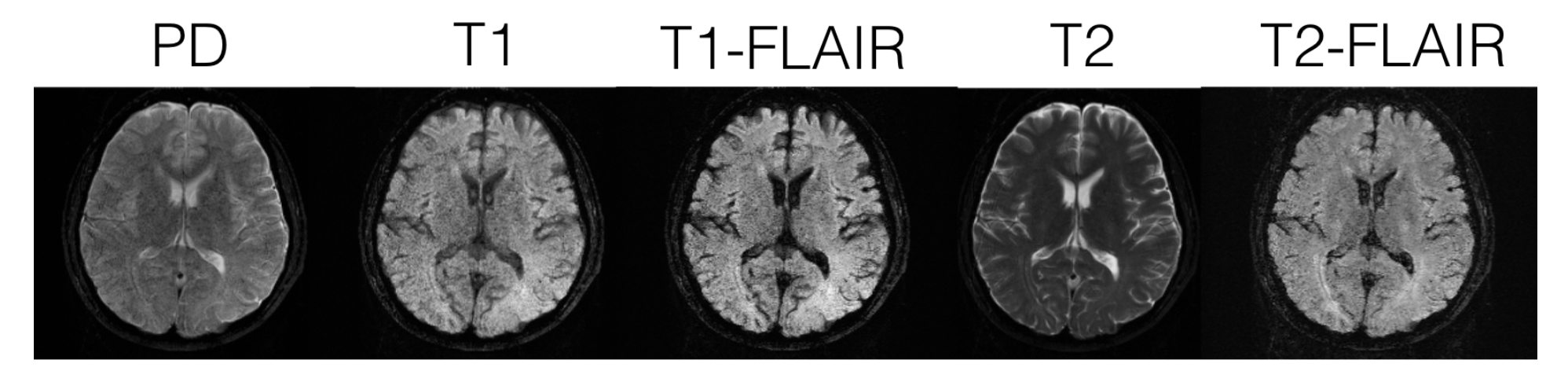
Figures 3a and 3b demonstrates the high-quality T1 and T2 maps reconstructed from the IR-prepared ES-GRASE data using parametric POCSMUSE method. Table 1a and 1b show the measured T1 and T2 values within selected regions of white matter (WM) and gray matter (GM) are very close to the ground truth. Figure 4 demonstrates the synthetic PDW, T1W, T1-FLAIR, T2W, and T2-FLAIR images generated from the measured T1 and T2 maps, in which the CSF signal is successfully eliminated for FLAIR-contrast images.Discussion and conclusions
Here we report a simultaneous T1 and T2 mapping method enabled by IR-prepared ES-GRASE scan and parametric POCSMUSE reconstruction. The data for parametric mapping can be obtained within 5 seconds for one slice, in which unwanted echoes are reduced and the parametric maps can be accurately measured by parametric POCSMUSE. The new method can also generate a complete set of high-quality synthetic multi-contrast images from the measured parametric maps, enabling multi-contrast imaging that is feasible for clinical settings. Experimental results demonstrate the successful performance of this new technology.Acknowledgements
No acknowledgement found.References
1. Hennig J. Echoes—how to generate, recognize, use or avoid them in MR imaging sequences. I. Fundamental and not so fundamental properties of spin echoes. Concepts Magn Reson 1991;3:125–143.
2. Chu ML, et al. A Single-Shot T2 Mapping Protocol Based on Echo-Split Gradient-Spin-Echo Acquisition and Parametric Multiplexed Sensitivity Encoding Based on Projection Onto Convex Sets Reconstruction. Magn Reson Med 2017; doi: 10.1002/mrm.26696.
3. Chen NK, et al. A robust multi-shot scan strategy for high-resolution diffusion-weighted MRI enabled by multiplexed sensitivity-encoding (MUSE). NeuroImage 2013;72:41–47.
Figures