5639
Single-shot multi-slice T1 mapping using inversion recovery radial FLASH and model-based reconstruction1Biomedizinische NMR Forschungs GmbH am Max-Planck-Institut für biophysikalische Chemie, Göttingen, Germany, 2Department of Interventional and Diagnostic Radiology of the University Medical Center Göttingen, Göttingen, Germany
Synopsis
Fast quantitative T1 mapping can be achieved within a single inversion recovery based on recent advances in real-time MRI and/or model-based reconstructions. However, efforts have been mainly focused on single-slice T1 mapping. To further take advantage of inherent data redundancy, we propose a single-shot high-resolution multi-slice T1 mapping technique which bases on a spoke-interleaved radial FLASH data acquisition scheme and a recent proposed model-based reconstruction technique. Initial results show that we could achieve high resolution seven-slice human brain T1 maps or three-slice abdominal T1 maps within 4 seconds.
Introduction
Recent advances in real-time MRI [1,2] and model-based reconstructions [3,4] enable fast quantitative T1 mapping within a single inversion recovery. However, efforts have been mainly focused on single-slice T1 mapping. To take full advantage of inherent data redundancy, we propose to combine an inversion-recovery spoke-interleaved radial FLASH data acquisition scheme with a model-based reconstruction technique for high-resolution multi-slice T1 mapping.
Methods/subjects:
Data acquisition starts with a single non-selective inversion pulse, followed by continuous spoke-interleaved data acquisition. The inter-slice distance is chosen to be equal the slice thickness. The relaxation process for each individual slice then follows the three-parameter model [5]:
$$M(t) = M_{ss} - (M_{ss} + M_{0})\cdot e^{-t\cdot R_{1}^{*}}$$ with $$$M_{ss}$$$ the steady-state magnetization, $$$M_{0}$$$ the equilibrium magnetization, and $$$ R_{1}^{*}$$$ the effective relaxation rate given by $$$R_{1}^{*} = \frac{1}{T_{1}} - \frac{\ln\cos(\alpha)}{n_{sl}\cdot TR}$$$, $$$n_{sl}$$$ is the number of slices. After data acquisition, all parameter maps and coil sensitivities are directly estimated from k-space using the sparsity-constrained model-based reconstruction technique [4]. The regularization parameters are chosen by visual inspection to obtain good SNR without compromising quantitative accuracy. All measurements were performed on a human 3T MRI system (Magnetom Prisma fit, Siemens Healthcare, Erlangen, Germany). The proposed method was experimentally validated with a commercial reference phantom (Diagnostic Sonar LTD, Scotland, UK) before in vivo applications. 4 subjects without known illness were then recruited among the students of the local University. Written informed consent according to the recommendations of the local ethics committee was obtained from all subjects prior to MRI. Phantom and brain studies were conducted with a standard 64-channel head coil, while abdominal scans were performed with an 18-element thorax coil in conjunction with 18 elements of the 32-element spine coil. Abdominal measurements were performed during a brief breath hold.Results
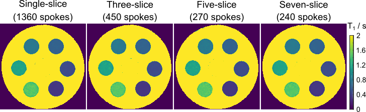
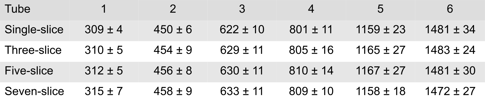
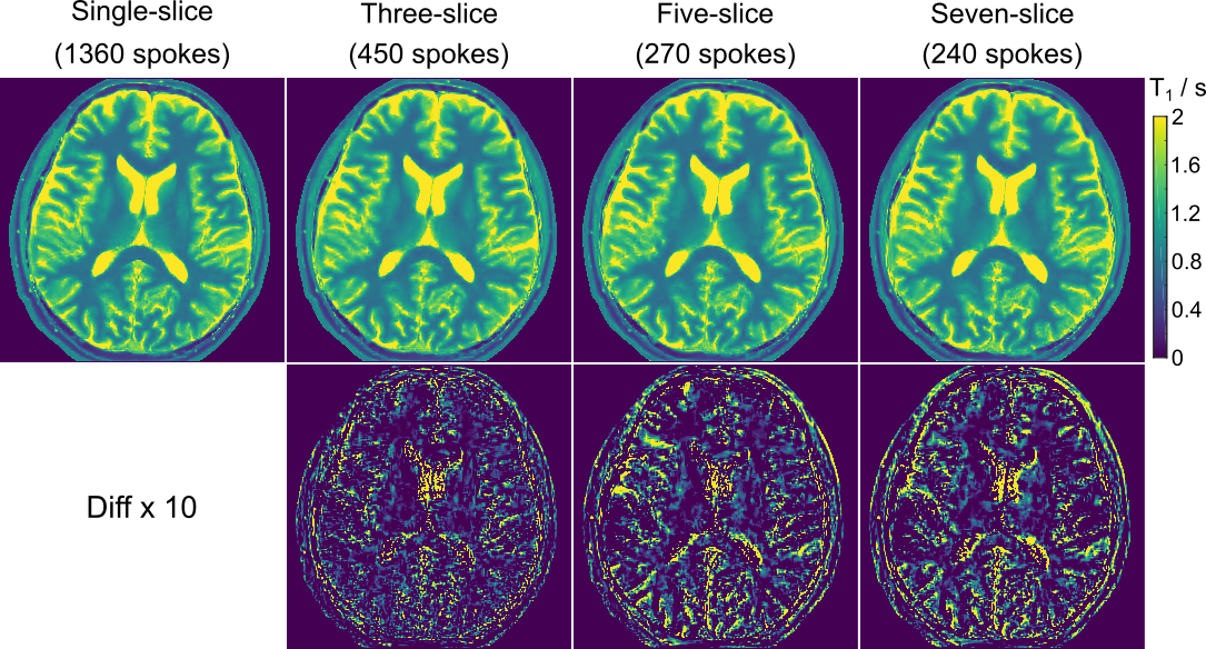
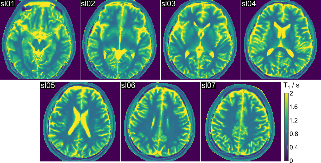
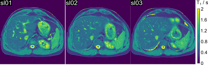
Figure 1 shows estimated phantom T1 maps (center-slice section) obtained from single, three, five and seven-slice acquisitions. Acquisition time was within 4 seconds in all cases. Visual inspection demonstrates marginal differences. This finding is quantitatively confirmed by ROI analyses summarized in Table 1. Figure 2 compares T1 maps (center-slice section) of the human brain. When increasing the number of slices, map quality remains good although a trend of increased differences in the T1 maps is visible. Figure 3 shows high-resolution (0.75 x 0.75 x 4 mm3) T1 maps (all slice sections) acquired within 4 seconds. A similar quality is achieved for applications to the human liver as depicted in Figure 4. In this case, all three maps (1.2 x 1.2 x 6 mm3) are estimated from datasets acquired with a 4 s breath hold.Discussion
In summary, we proposed a novel method which combines inversion recovery radial FLASH and model-based reconstruction for single-shot high-resolution multi-slice T1 mapping. Initial results show that we could achieve simultaneous 7-slice brain T1 maps or 3-slice abdominal T1 maps within a 4-second data acquisition. For further acceleration, the proposed method can also be combined with a multi-band acquisition [6].Acknowledgements
No acknowledgement found.References
[1]. Uecker M, Zhang S, Voit D, Karaus A, Merboldt KD, Frahm J. RealtimeMRI at a resolution of 20 ms. NMR Biomed 2010;23:986–994.
[2]. Wang X, Joseph AA, Kalentev O, Merboldt KD, Voit D, Roeloffs V, van Zalk M, Frahm J. High-resolution myocardial T1 mapping using single-shot inversion-recovery fast low-angle shot MRI with radial undersampling and iterative reconstruction. Br J Radiol 2016;89:20160255.
[3]. Tran-Gia J, Staeb D, Wech T, Hahn F, Koestler H. Model-based Acceleration of Parameter mapping (MAP) for saturation prepared radially acquired data. Magn Reson Med 2013;70:1524–1534.
[4]. Wang X, Roeloffs V, Klosowski J, Tan Z, Voit D, Uecker M and Frahm J. Model-based T1 mapping with sparsity constraints using single-shot inversion-recovery radial FLASH. Magn Reson Med, DOI: 10.1002/mrm.26726 (2017).
[5]. Look DC, Locker DR. Time saving in measurement of NMR and EPRrelaxation times. Rev Sci Instrum 1970;41:250–251.
[6]. Rosenzweig S, Holme H, Wilke R, Voit D, Frahm J, Uecker M, Simultaneous Multi-Slice Reconstruction Using Regularized Nonlinear Inversion: SMS-NLINV, Magn Reson Med, DOI:10.1002/mrm.26878 (2017).
Figures