5613
Brain Tumor and Prostate EPI MR Elastography Distortion Reduction and Correction Using rFOV and PSF-EPI Techniques1Radiology, Mayo Clinic, Rochester, MN, United States
Synopsis
Image distortion is a common problem in EPI-based MR elastography (MRE) acquisitions, especially in regions with high susceptibility, including regions adjacent to air-tissue interfaces, such as the paranasal sinuses and the prostate. In this study, we explored a reduced-field-of-view (rFOV) technique and a point-spread-function (PSF) mapping EPI correction method to reduce or correct the distortions in EPI MRE images of brain tumors and the prostate.
Introduction
EPI-based MR elastography (MRE) acquisitions are prone to image distortion caused by local susceptibility artifacts. It is especially challenging in regions with high susceptibility, including adjacent to air-tissue interfaces such as the paranasal sinuses and the prostate (1). These distortions may affect the accuracy of stiffness measurements in these small regions. Many approaches have been developed to reduce or correct the EPI distortion. For instance, reduced-field-of-view (rFOV) EPI using 2D-RF excitations can effectively reduce the distortion by limiting the imaging FOV to a targeted region, allowing for a reduction in the EPI readout train (2), and point-spread-function echo-planar image (PSF-EPI) mapping can be performed as a calibration for EPI distortion correction (3,4). In this study, we implemented rFOV and PSF-EPI techniques into brain tumor and prostate MRE protocols and evaluated how well each technique reduced or corrected the EPI distortions.Methods
Under an IRB-approved protocol, 5 patients with brain tumors were scanned on a recently developed, compact, 3T system with high-performance gradients, capable of 80 mT/m gradients and 700 T/m/s slew rate (5), and an 8-channel receiver coil (Invivo, Gainesville, FL). B0 and linear concomitant fields from the asymmetric gradients are compensated by frequency tracking (6) and gradient pre-emphasis (7), respectively. Three MRE experiments were conducted for comparison. First, a standard 2-mm-isotropic EPI MRE with 80-Hz mechanical vibrations; TR/TE=2976/60.9 ms; FOV=24 cm; 120×120 matrix; 34 contiguous 2-mm-thick axial slices; 2x ASSET acceleration; 316 μs echo spacing, 4 phase offsets; 4 averages and total acquisition time of 4:51 minutes. Second, a PSF-EPI scan to collect the calibration data with matched imaging parameters to the standard MRE except for TR/TE=2000/26.6 ms and a scan time of 80 seconds. A correction kernel calculated from the PSF-EPI approach was applied to the standard MRE data for distortion correction as in (3). Third, rFOV-MRE with a 1/3 FOV in the phase-encoding direction was used to reduce the distortion using a 120×40 matrix, no ASSET acceleration, and with the other parameters matched to the standard brain MRE.
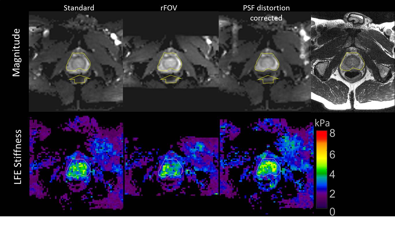
Six normal prostate volunteers were scanned on a whole-body, 3T, GE scanner with an 8-channel torso coil. Three scans were conducted. First, a standard 3-mm-isotropic EPI-MRE with 90-Hz vibrations induced by three rigid, passive, drivers placed around the pelvis; TR/TE=2000/80.3 ms; FOV=38.4 cm; 128×128 matrix; 3-mm-thick axial slices; 2x ASSET acceleration; 544 μs echo spacing, 8 offsets; an acquisition time of 4:20 minutes. Second, a PSF-EPI scan matched to the standard MRE except for TR/TE=1800/46.4 ms and a scan time of 58 seconds. Third, rFOV-MRE with 1/3 FOV, 128×44 matrix, and other parameters matched to the standard prostate MRE.
Results
A representative patient with a meningioma adjacent to the maxillary sinus is presented in Figure 1. A significant amount of image distortion arising from the large susceptibility gradient can be seen on the magnitude image for the standard MRE. The distortion was reduced by using the rFOV technique, and substantially corrected by the PSF-based distortion correction method. This tumor stiffness was measured as 1.7±0.6, 2.0±0.9, and 2.1±0.9 kPa using the standard EPI-MRE, rFOV, and distortion-correction approaches, respectively. Figure 2 shows a representative case of prostate MRE in a healthy volunteer. Similar to brain MRE, the geometric distortion was reduced using rFOV-MRE, and was corrected by the PSF-correction method. The corresponding stiffness values measured in the prostate ROI were 3.2±0.8, 3.0±0.8, and 3.6±0.9 kPa, with the standard EPI-MRE, rFOV-MRE, and PSF-corrected-EPI-MRE techniques, respectively.Discussion and Conclusion
As demonstrated in these brain tumor and prostate applications, the geometric distortion in the standard EPI-MRE images can be reduced using rFOV-MRE techniques, and can be corrected using the PSF method. Different from the techniques using B0 maps, or the one used in the FSL-TOPUP tool (8), PSF-EPI uses an extra PSF phase-encoding step (similar to spin-echo phase-encoding) before the EPI phase blips. It is able to simultaneously acquire a set of images with and without EPI distortions, which can be used to generate the distortion-correction kernel for MRE images. In the future, we will look at integrating the PSF method into the current rFOV-MRE sequence by adding another EPI train. This combination could lead to a more efficient approach for the MRE distortion correction. The difference in the stiffness measurements from the three experiments may be caused by the wave field changes before and after the reduction or correction of the EPI distortion. Given the stiffness calculation is sensitive to the SNR, this difference may also be, in part, a result from the smoothing effects of geometric correction, which lead to an SNR increase. Further investigation is needed to confirm the reduced distortion and increased SNR lead to improved accuracy of the tissue stiffness measurements.Acknowledgements
This work was supported by the grants from the National Institute of Health RO1 EB001981 and U01 EB024450.References
- Glaser KJ, Manduca A, Ehman RL. Review of MR elastography applications and recent developments. J Magn Reson Imaging 2012;36(4):757-774.
- Sui Y, Forghanian-Arani A, Joshua D. Trzasko, Arunachalam SP, Glaser KJ, Lake DS, McGee KP, Manduca A, Rossman PJ, Ehman RL, Araoz aPA. Improvements in Cardiac MR Elastography Using Reduced FOV Techniques. ISMRM 2016:2880.
- In MH, Speck O. Highly accelerated PSF-mapping for EPI distortion correction with improved fidelity. MAGMA 2012;25(3):183-192.
- In MH, Posnansky O, Speck O. High-resolution distortion-free diffusion imaging using hybrid spin-warp and echo-planar PSF-encoding approach. Neuroimage 2017;148:20-30.
- Weavers PT, Shu Y, Tao S, Huston J, Lee S-K, Graziani D, Mathieu J-B, Trzasko JD, Foo TKF, Bernstein MA. Technical Note: Compact three-tesla magnetic resonance imager with high-performance gradients passes ACR image quality and acoustic noise tests. Medical Physics 2016;43(3):1259-1264.
- Weavers PT, Tao S, Trzasko JD, Frigo LM, Shu Y, Frick MA, Lee SK, Foo TK, Bernstein MA. B0 concomitant field compensation for MRI systems employing asymmetric transverse gradient coils. Magn Reson Med 2017.
- Tao SZ, Weavers PT, Trzasko JD, Shu YH, Huston J, Lee SK, Frigo LM, Bernstein MA. Gradient Pre-Emphasis to Counteract First-Order Concomitant Fields on Asymmetric MRI Gradient Systems. Magnetic Resonance in Medicine 2017;77(6):2250-2262.
- Fehlner A, Hirsch S, Weygandt M, Christophel T, Barnhill E, Kadobianskyi M, Braun J, Bernarding J, Lutzkendorf R, Sack I, Hetzer S. Increasing the spatial resolution and sensitivity of magnetic resonance elastography by correcting for subject motion and susceptibility-induced image distortions. J Magn Reson Imaging 2017;46(1):134-141.
Figures