5610
Robust Water-Fat Separation in Multi-Echo GRE Sequence using Patch-Based Neural Network1Korea Advanced Institute of Science and Technology (KAIST), DaeJeon, Republic of Korea
Synopsis
The water-fat separation techniques using a multi-echo GRE sequence has suffered from an inaccurate and swapped water-fat separation results caused by several issues. In the abstract, we propose a robust water-fat separation method using patch-based neural network to overcome this problem. The neural network is trained using the relationship between the multi-echo images obtained from the multi-echo GRE sequence and the reliable water-fat separated images that are reconstructed by IDEAL from the multiple single-echo GRE acquisitions with different echo times. The in-vivo experiment results show the proposed method can successfully separate accurate water-fat images from the multi-echo GRE images in comparison with IDEAL.
Introduction
Method
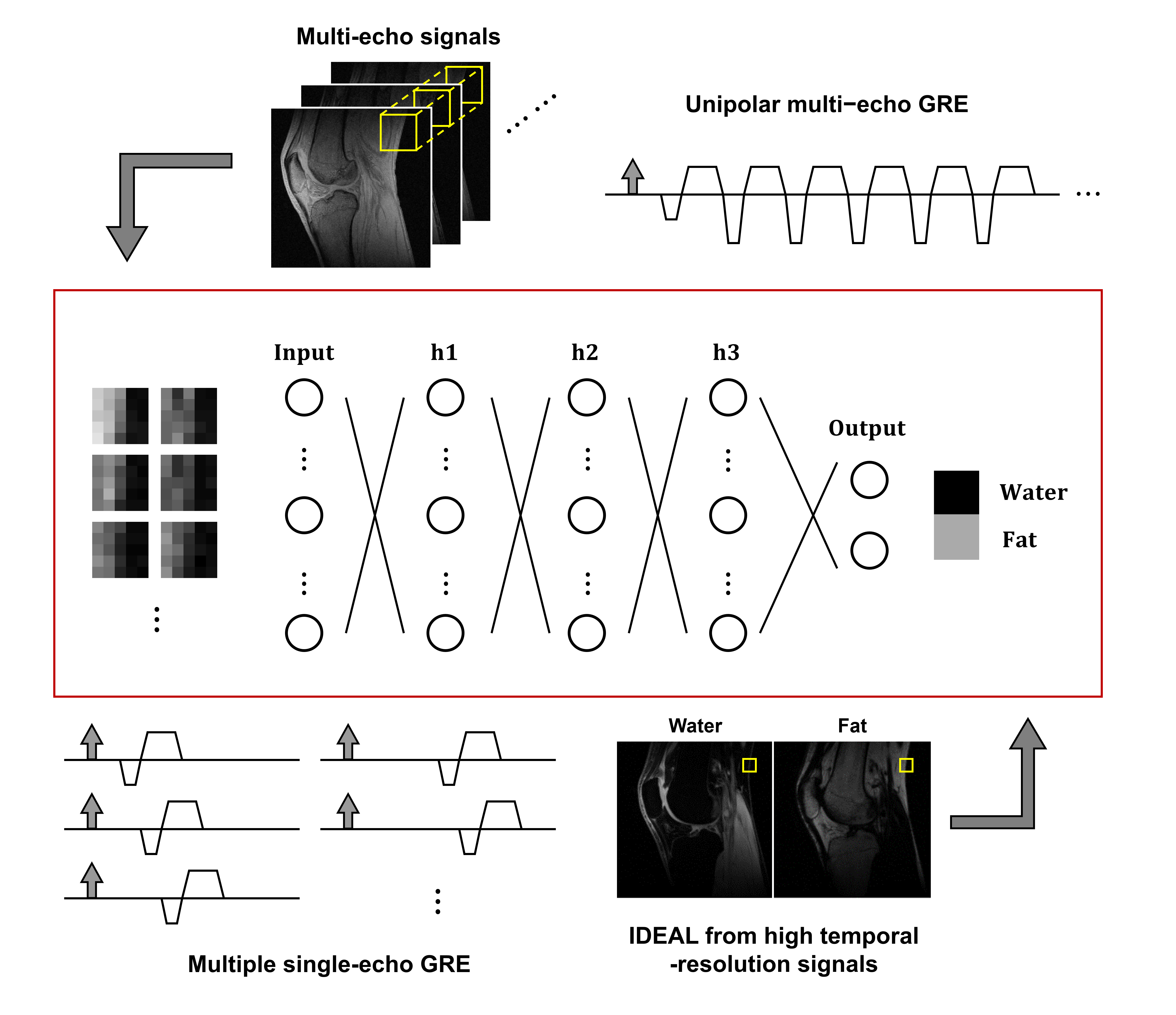
Figure 1 shows overall scheme of the proposed neural network. The proposed method trains the network using the relationship between the multi-echo images from the multi-echo GRE sequence and the reliable water-fat separated images. Then, multi-echo images are the input data to the trained neural network and the accurate water-fat separated images could be obtained as outputs. It is assumed that the reliable water-fat separated images can be obtained by the IDEAL from the multiple single-echo GRE acquisitions with different echo times. The multiple single-echo GRE acquisitions can eliminate the issues from the eddy current and other system imperfections and achieve a short echo-spacing time. The input and output of the proposed neural network are the bunch of patches of the multi-echo images and the separated water-fat signals, respectively. The previous water-fat separation methods including Dixon method and iterative decomposition of water and fat with echo asymmetry and least-squares estimation (IDEAL) fundamentally perform the pixel-by-pixel operation.5,6 The patch-based method utilizes the nearby voxels so that it can provide an additional information such as distorted phase or chemical shift, and prevent the water-fat images from the local minima. Furthermore, the patch-based implementation can easily obtain sufficient training sets from a few data acquisitions.
The experiments were conducted on a KAIST 3T MRI system (Verio, Siemens Medical Solutions, Inc., Erlangen, Germany). The unipolar multi-echo GRE sequence was implemented using eight-channel knee coil with following parameters: TR=20ms; 1st echo time=2.03ms; echo-spacing time=2.89ms; the number of echoes=6; slice thickness=5mm; FOV=192mmⅹ192mm; and matrix size=192ⅹ192. The high temporal-resolution images from the multiple single-echo GRE acquisitions were obtained using eight-channel knee coil with following parameters: TR=18ms; 1st echo time=3.30ms; echo-spacing time=0.25ms; the number of echoes=25; slice thickness=5mm; FOV=192mmⅹ192mm; and matrix size=192ⅹ192. 16 slices in coronal and sagittal views were obtained, respectively. For each view, 15 slices were exploited for the training and 1 slice was exploited to validate the neural network. The IDEAL with the region growing scheme was implemented using the ISMRM fat-water toolbox.3,7 The neural network was constructed using the TensorFlow.8 The input of the neural network was 5ⅹ5 patches of six echoes and the output was the separated water-fat signals at the center point of the patches. The patches were generated separately for each channel of the receiver RF coil, so that the number of training and validation patches were about 9.1M and 0.3M, respectively. The proposed neural network included fully connected three hidden layers, each of which had 300 neurons. The learning rate, batch size, and the number of training epochs were 0.005, 2000, and 1000, respectively. The Adam optimizer and mean-squared loss were used to train the neural network.
Results
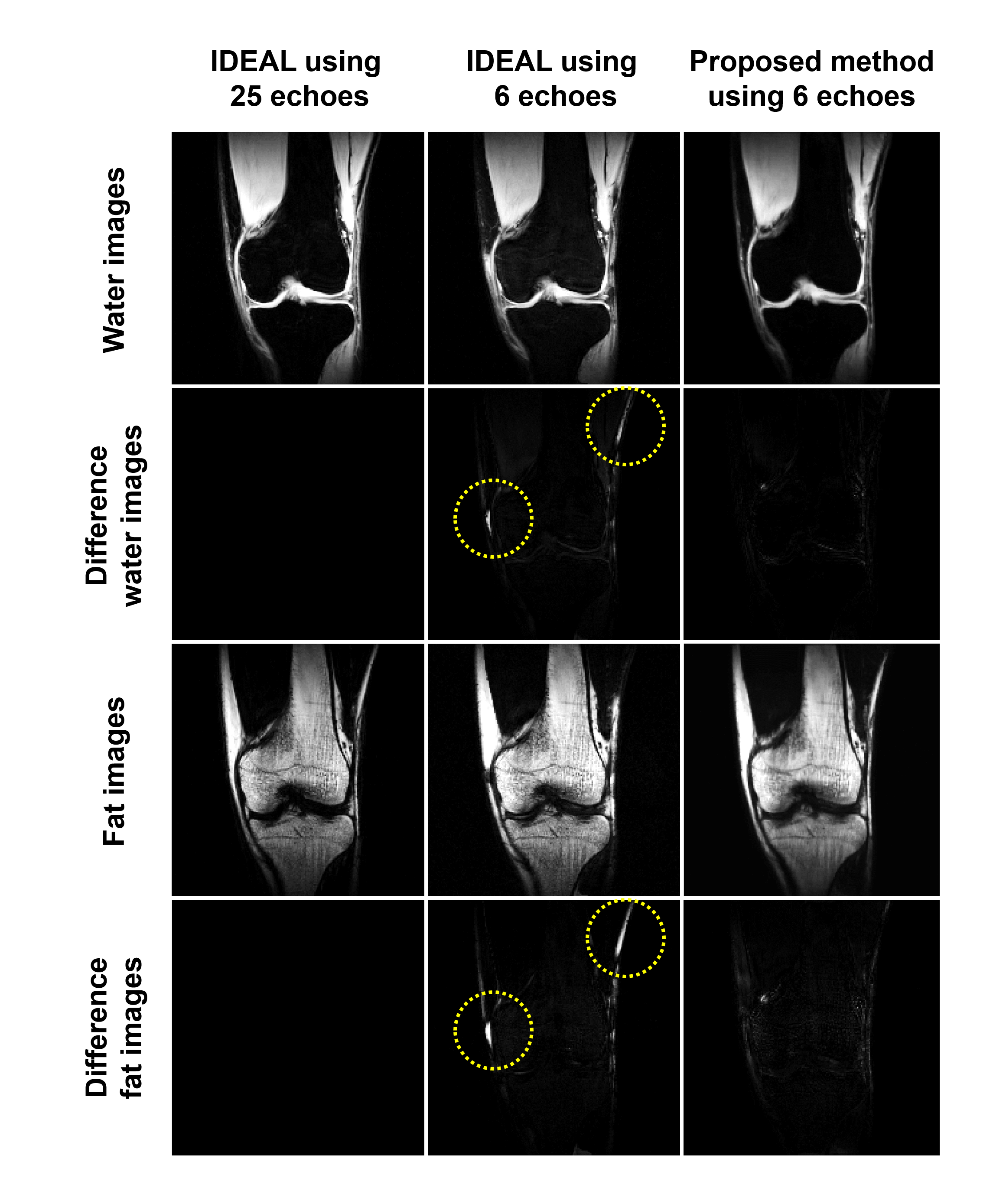
Figure 2 shows the experiment results of the validation image. The left column presents the reference IDEAL results using 25 echoes obtained from the multiple single-echo GRE acquisitions. The middle and right columns present the results of IDEAL and the proposed method using six echoes from the multi-echo GRE sequence. The second and fourth rows show the difference from the reference results. The regions of yellow circles show inaccurate swapped water-fat results of IDEAL from the six echoes. The proposed method successfully separates accurate water-fat images.Discussion and Conclusion
The experiment results demonstrates that the proposed method can separate accurate water-fat images using the multi-echo GRE sequence. The proposed method inputs the multi-echo image patches to the neural network and obtained the water-fat separated images. The patch-based implementation can increase the number of the training set and bring the flexibility of input image size. However, the patch-based implementation can cause a blurring artifact in the water-fat separated images. The expansion of the neural network which inputs the entire image and the estimation of quantitative data such as field map and R2* map are the further works of this abstract.Acknowledgements
This research was partly supported by Institute for Information & communications Technology Promotion (IITP) grant funded by the Korea government (MSIT) (No.2017-0-01778, Development of Explainable Human-level Deep Machine Learning Inference Framework), and by the Ministry of Health & Welfare, Republic of Korea (grant number : HI14C1135). We acknowledge the use of the Fat-Water Toolbox (http://ismrm.org/workshops/FatWater12/data.htm) for some of the results shown in this article.References
1. Yu H, Shimakawa A, McKenzie CA, Brodsky E, Brittain JH, Reeder SB. Multiecho water-fat separation and simultaneous R2* estimation with multifrequency fat spectrum modeling. Magn. Reson. Med. 2008;60:1122–1134.
2. Yu H, Shimakawa A, McKenzie CA, Lu W, Reeder SB, Hinks RS, Brittain JH. Phase and amplitude correction for multi-echo water-fat separation with bipolar acquisitions. J. Magn. Reson. Imaging 2010;31:1264–1271.
3. Yu H, Reeder SB, Shimakawa A, Brittain JH, Pelc NJ. Field map estimation with a region growing scheme for iterative 3-point water-fat decomposition. Magn. Reson. Med. 2005;54:1032–1039.
4. Hernando D, Kellman P, Haldar JP, Liang Z-P. Robust water/fat separation in the presence of large field inhomogeneities using a graph cut algorithm. Magn. Reson. Med. 2009:79–90.
5. Dixon WT. Simple proton spectroscopic imaging. Radiology 1984;153:189–194.
6. Reeder SB, Pineda AR, Wen Z, Shimakawa A, Yu H, Brittain JH, Gold GE, Beaulieu CH, Pelc NJ. Iterative decomposition of water and fat with echo asymmetry and least-squares estimation (IDEAL): application with fast spin-echo imaging. Magn. Reson. Med. 2005;54:636–644.
7. Hu HH, Börnert P, Hernando D, Kellman P, Ma J, Reeder S, Sirlin C. ISMRM workshop on fat-water separation: insights, applications and progress in MRI. Magn. Reson. Med. 2012;68:378–388.
8. Abadi M. TensorFlow: learning functions at scale. ACM SIGPLAN Notices 2016;51:1–1.
Figures