5604
Improve PCASL Brain Imaging at 7T Using Dynamically Applied B1+ Shimming Solutions1Center for Magnetic Resonance Research, School of Medicine, University of Minnesota, Minneapolis, MN, United States
Synopsis
The rapid decline of transmit B1 ( B1+) toward the inferior brain regions and large B1+ inhomogeneity across blood feeding arteries at the labeling plane impose great challenges for high quality pseudo-continuous arterial spin labeling (PCASL) brain imaging at 7T. Recent studies at 7T have suggested that dynamically applied B1+ shimming solutions separately targeted to imaging and labeling regions can help to overcome these challenges. We implemented and evaluated the dynamic B1+ shimming approach for PCASL brain imaging at 7T, demonstrating that such an approach can greatly improve B1+ performance for blood tagging.
Purpose
Because of increases in blood T1 1 and signal-to-noise ratio (SNR), 7T should benefit arterial spin labeling (ASL) imaging, and may improve its ability and overcome challenges at 3T in clinical research studies of diseases having dramatically prolonged arterial transit time, such as stroke. However, to date, few 7T studies 2-5 have shown promising results using either pulsed arterial spin labeling (PASL), such as FAIR 6, or pseudo-continuous arterial spin labeling (PCASL) imaging methods 7,8, primarily because the expected benefits cannot be fully realized until the technical challenges at 7T have been overcome. The limited transmit B1 (B1+) coverage of typical head coils cannot provide sufficient temporal bolus duration for PASL methods. The result is not only low perfusion SNR but also a reduction in the reliability of cerebral blood flow (CBF) quantification. Although PCASL can overcome such limitations by tagging inflowing blood within the labeling plane overtime, the rapidly decreasing B1+ toward the inferior brain regions can dramatically decrease labeling efficiency while large B1+ inhomogeneities across blood feeding arteries can result in significantly varied labeling efficiencies for different perfusion territories thus complicating CBF quantification 5.
Recent studies at 7T have suggested that dynamically applied B1+ shimming solutions separately targeted to imaging and labeling regions can help to overcome the B1+ challenges and improve ASL imaging 3. We implemented and evaluated the dynamic B1+ shimming approach for PCASL brain imaging at 7T, demonstrating that such an approach can greatly improve B1+ performance for blood tagging.
Methods
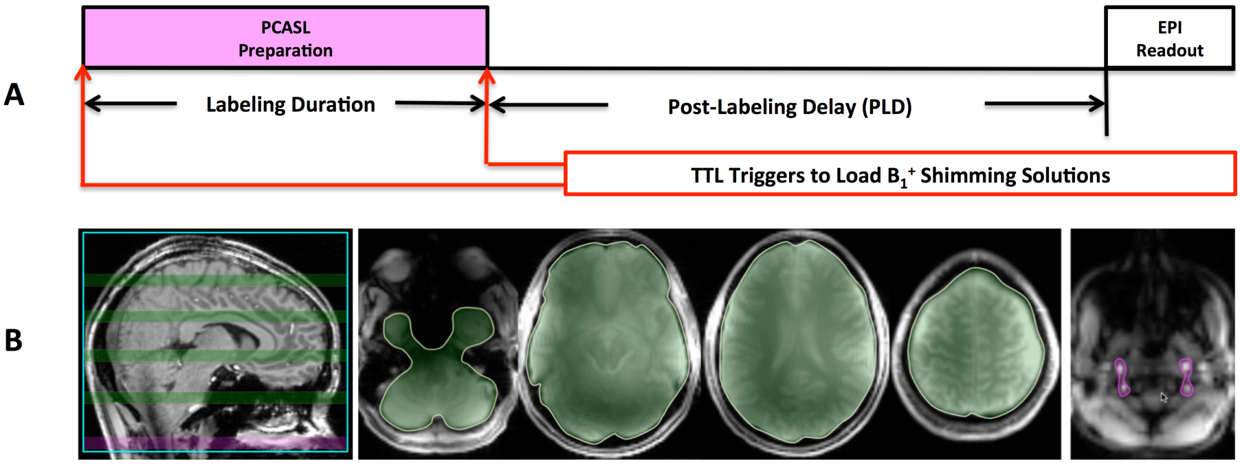
Healthy volunteers were imaged for this study after having provided written consent to participate in an IRB approved protocol. Studies were performed on Siemens 7T whole body MRI scanner and using a Nova head coil with 32 channels for signal reception and 8 channels for RF transmission. The B1+ optimizations in this study utilized a phase-only B1+ shimming approach based on the data acquired from a single small flip angle calibration scan consisting of imaging slices covering the target regions 9,10. The dynamic B1+ shimming strategy consisted of 2 solutions: one for the whole brain which provides a circular polarization style B1+ field with a Gaussian spatial distribution (denoted as CP shimming method), and another for blood tagging based on a tradeoff between B1+ homogeneity and RF efficiency (denoted as tradeoff shimming method). The sequence diagram for PCASL imaging with dynamically applied B1+ shimming solutions is presented in Figure 1A, and the regions of interests (ROIs) for B1+ shimming solutions for the whole brain and blood tagging are shown in Figure 1B. To evaluate the B1+ performance resulting from the different B1+ shimming solutions, B1+ maps were obtained using 3D actual flip angle imaging (AFI) 11. To address B0 issues, 3rd order shimming was performed over a volume covering both imaging slices and labeling plane (Figure 1B) 5.
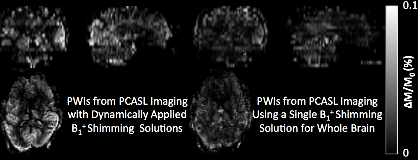
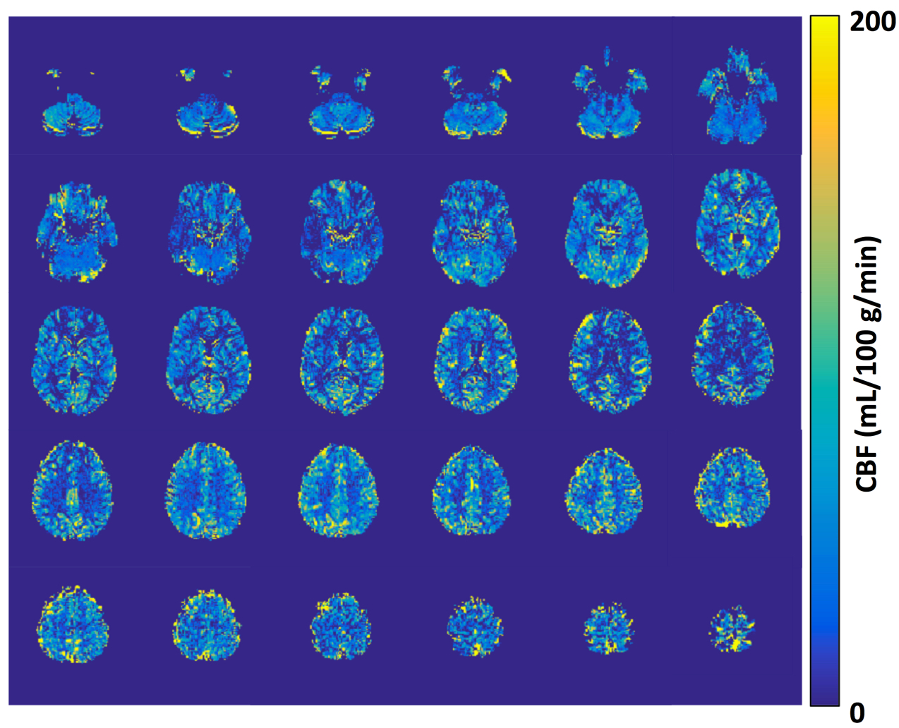
Echo planar imaging was used as the image readout for multi-slice PCASL acquisitions with a 1.5 s labeling duration and 1.6 or 2.0 s post-labeling delays (PLDs). A comparison study was first performed to evaluate the results from low-resolution PCASL imaging with and without dynamically applied B1+ shimming solutions (Figure 3). Once it was confirmed that high quality low-resolution perfusion maps were achieved with the improved B1+ performance for blood tagging, high-resolution PCASL imaging with dynamically applied B1+ shimming solutions was further performed (Figure 5).
Post-processing was performed with SPM. The CBF quantification employed the single-blood compartment model 12 and in-house MATLAB scripts. Statistical analyses were performed using the GraphPad.
Results and Discussions
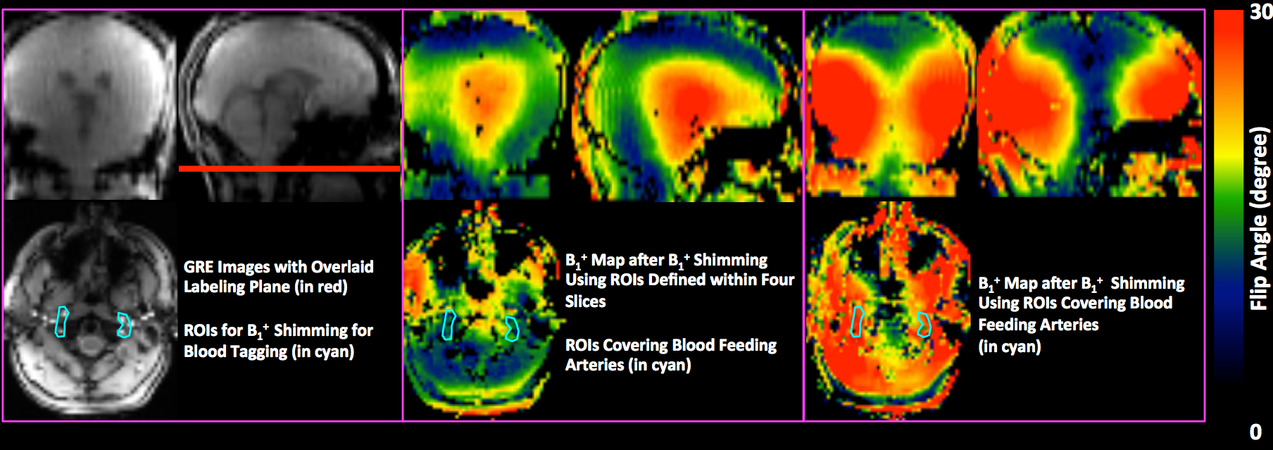
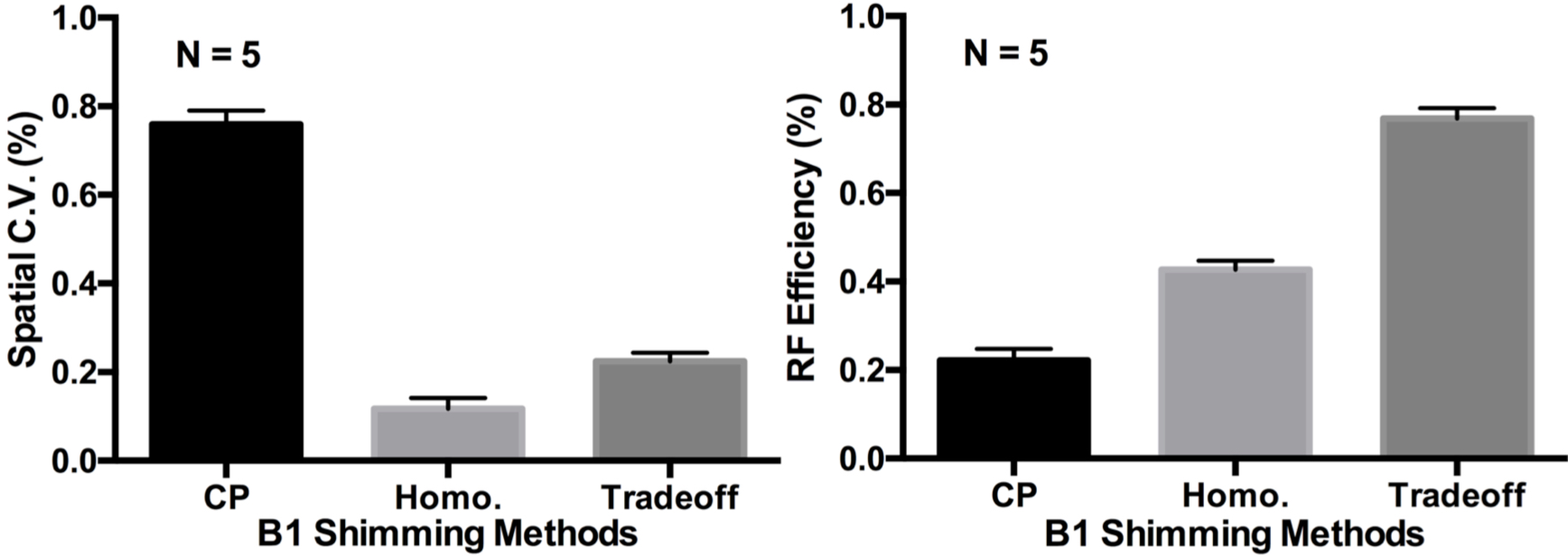
Our study shows that both the B1+ homogeneity and RF efficiency can be greatly improved for blood tagging by applying a tradeoff B1+ shimming solution based on ROIs covering only the blood feeding arteries at the level of the labeling plane. Figure 2 shows the B1+ maps from one typical subject at the labeling plane after B1+ optimization for the whole brain using the CP shimming method and for blood tagging using the tradeoff shimming method. The normalized perfusion-weighted images (PWIs) of one subject from the comparison study are presented in Figure 3. Quantitative analysis results further suggest that the tradeoff B1+ shimming method provides a reasonable balance between B1+ spatial variability and RF efficiency (Figure 4). High quality high-resolution CBF maps from PCASL imaging with dynamically applied B1+ shimming solutions have been successfully obtained from subjects (Figure 5). Further technical development, such as using slice specific RF shimming 13, is needed to improve B1+ performance in the imaging region.Conclusions
The dynamic application of B1+ shimming solutions separately targeted to imaging and labeling regions can significantly improve PCASL brain imaging at 7T.Acknowledgements
P41 EB015894, P30 NS076408, UMF0003900, and UL1TR000114. The content is solely the responsibility of the authors and does not necessarily represent the official views of the National Institutes of Health.References
1. Zhang X, Petersen ET, Ghariq E, et al. In vivo blood T(1) measurements at 1.5 T, 3 T, and 7 T. Magn Reson Med. 2013;70(4):1082-1086.
2. Luh WM, Talagala SL, Li TQ, Bandettini PA. Pseudo-continuous arterial spin labeling at 7 T for human brain: estimation and correction for off-resonance effects using a Prescan. Magn Reson Med. 2013;69(2):402-410.
3. Li X, Auerbach EJ, Van de Moortele PF, Ugurbil K, Metzger GJ. Quantitative single breath-hold renal arterial spin labeling imaging at 7T. Magn Reson Med. 2017.
4. Zuo Z, Wang R, Zhuo Y, Xue R, St Lawrence KS, Wang DJ. Turbo-FLASH based arterial spin labeled perfusion MRI at 7 T. PLoS One. 2013;8(6):e66612.
5. Li X, Wang, D, Moeller, S, Ugurbil, K, & Metzger, GJ. Feasibility of Applying MB EPI pCASL for High-Resolution Whole Brain Perfusion Imaging at 7T. In: Proceedings of the 21st Annual Meeting of ISMRM, Milano, Italy, 2014:Abstract 0995.
6. Kim SG. Quantification of relative cerebral blood flow change by flow-sensitive alternating inversion recovery (FAIR) technique: application to functional mapping. Magn Reson Med. 1995;34(3):293-301.
7. Dai W, Garcia D, de Bazelaire C, Alsop DC. Continuous flow-driven inversion for arterial spin labeling using pulsed radio frequency and gradient fields. Magn Reson Med. 2008;60(6):1488-1497.
8. Wu WC, Fernandez-Seara M, Detre JA, Wehrli FW, Wang J. A theoretical and experimental investigation of the tagging efficiency of pseudocontinuous arterial spin labeling. Magn Reson Med. 2007;58(5):1020-1027.
9. Van de Moortele PF, Akgun C, Adriany G, et al. B(1) destructive interferences and spatial phase patterns at 7 T with a head transceiver array coil. Magn Reson Med. 2005;54(6):1503-1518.
10. Metzger GJ, Auerbach EJ, Akgun C, et al. Dynamically applied B1+ shimming solutions for non-contrast enhanced renal angiography at 7.0 Tesla. Magn Reson Med. 2013;69(1):114-126.
11. Yarnykh VL. Actual flip-angle imaging in the pulsed steady state: a method for rapid three-dimensional mapping of the transmitted radiofrequency field. Magn Reson Med. 2007;57(1):192-200.
12. Alsop DC, Detre JA, Golay X, et al. Recommended implementation of arterial spin-labeled perfusion MRI for clinical applications: A consensus of the ISMRM perfusion study group and the European consortium for ASL in dementia. Magn Reson Med. 2015;73(1):102-116.
13. Wu X, Schmitter S, Auerbach EJ, Ugurbil K, Van de Moortele PF. A generalized slab-wise framework for parallel transmit multiband RF pulse design. Magn Reson Med. Apr 2016;75(4):1444-1456.
Figures