5602
Real-time ECG-gated continuous vibration multi-shot spiral elastography acquisition: application to time-resolved cerebral stiffness measurements during arterial pulsation.1Department of Radiology, Charité - Universitätsmedizin Berlin, Berlin, Germany, 2Visage Imaging GmbH, Berlin, Germany, 3Institute for Medical Informatics, Charité - Universitätsmedizin Berlin, Berlin, Germany
Synopsis
We implemented a versatile spiral MRE sequence that synchronizes the acquisition in real-time to a continuous harmonic vibration. Using multi-shot gradient echo readout and fractional motion encoding, multiple phases can be acquired in the cardiac cycle with high temporal resolution. Optionally, prospective respiratory gating and slice tracking allow for imaging moving organs like the heart or the liver in free breathing. As a first application, we measured cerebral stiffness maps in multiple phases of the cardiac cycle to detect changes induced by arterial pulsation.
Introduction
Magnetic resonance elastography (MRE) is a robust technique for in vivo detection of tissue stiffness, but has limited temporal resolution. This particularly compromises MRE in moving organs such as the heart [1] or MRE applications for detecting stiffness variations in the brain during arterial pulsation [2]. To overcome this limitation we developed an ECG-gated spiral MRE acquisition technique that allows for purely continuous harmonic vibrations and respiration gating. MRE based on mechanical stimulation by continuous harmonic oscillations has been demonstrated as an important feature to avoid transient wave effects, to accelerate MRE by synchronizing single-shot image acquisition to steady-state oscillations and to use oscillating pressurized air as the driving force for mechanical actuators [3, 4]. Multi-shot spiral k-space readout minimizes TE and is thus a very time-efficient sampling scheme with intrinsic flow compensation [5]. In diffusion imaging, high-resolution images can be obtained this way [6]. In a first in-vivo experiment, we applied the new method in the brain to detect possible changes of tissue stiffness due to the arterial pulse wave.Methods
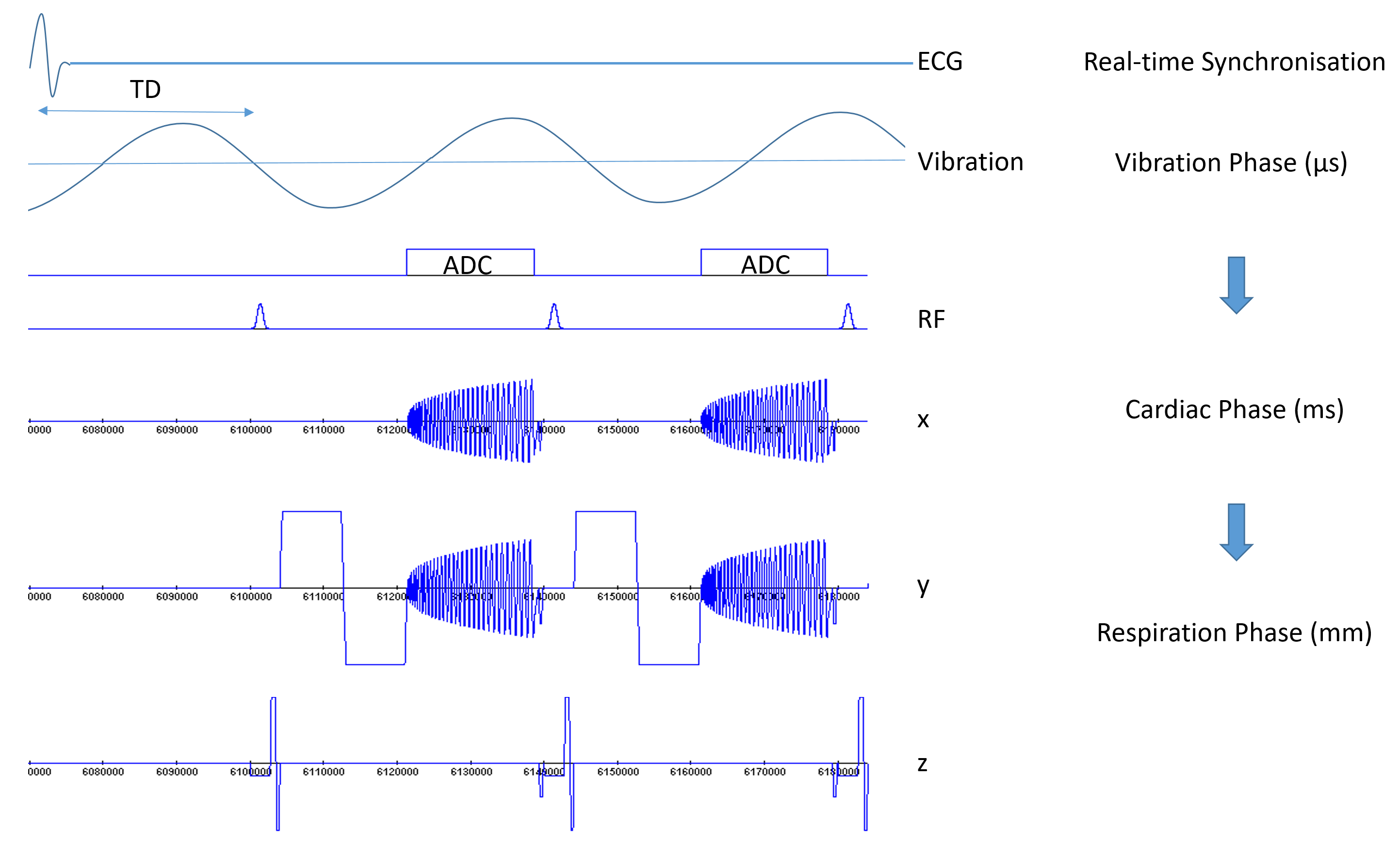
A sequence diagram is shown in figure 1. The new concept of continuous ECG-triggered MRE is based on the following hierarchy of external events: 1) the external harmonic motion has highest priority for sequence synchronization, i.e. start of motion encoding and image acquisition is exact on the microsecond scale due to clock synchronization of MRI scanner and vibration generator. 2) The cardiac phase was binned into intervals of 40 ms (a vibration period) in order to match the MRE acquisition to the ECG. 3) For future cardiac applications, we implemented the option of respiratory gating based on real-time pencil beam navigators. Overall, motion encoding and acquisition of a k-space segment was performed only at matching wave phases, during the right ECG phase and – if enabled – in the right respiration phase, e.g. during expiration. First experiments were performed in the brain of healthy volunteers. Pressurized air driven actuators excited the head continuously at a vibration frequency of 25 Hz. Using finger-pulse triggering, we acquired 12 phases in each cardiac cycle with TR = 40 ms temporal resolution, thus covering approx. half of a cardiac phase. We used a 15 ms bipolar motion-encoding gradient (MEG) giving an echo time (TE) of 20 ms. Readout time for one spiral trajectory was 17 ms. Ten shots resulted in a 192x192 image matrix of 300x300 mm² field-of-view. Eight motion-encoding steps, equally spaced over a wave period, required 240 heartbeats for acquisition of a single-slice wave field including all three encoding directions. Post processing was based on the tomoelastography pipeline proposed in [3,4].Results
A sample set of data acquired in the brain of a healthy volunteer is shown in figure 2. A significant change of brain stiffness (in terms of shear wave speed, in m/s) was observed during the acquisition window of 480 ms. The arterial peak systole occurred approx. during acquisition of the third image in this sequence of wave data. At this time, an increase of stiffness is visible in most parts of brain tissue in this image slice, which could be related to compression stiffening due to poroelastic properties of brain matter.Discussion
The implemented sequence is very flexible. It can be run in single-shot mode with low spatial and temporal resolution (due to the long readout) as well as in high-resolution mode using segmented multi-shot acquisition. It supports gradient echo as well as spin-echo readout and incorporates respiratory navigators with prospective respiratory gating and motion compensation in order to facilitate MRE acquisitions in other organs like the heart or the abdomen without the need for breath holding. In diffusion imaging with multi-shot spiral readout, a phase correction of the individual shots is essential. Without it, phase errors introduced by smallest bulk motions result in severe image artefacts. However, a phase correction requires an additional navigator readout before each k-space interleave. The additional time for this navigator adds to the echo time, thereby reducing the efficiency of the MRE scan. It is an encouraging result of this feasibility study that the image quality in MRE acquisitions appears to be sufficiently good even without this extra phase correction, at least for continuous harmonic vibration. Ongoing experiments in the brain will be compared to intrinsically activated MRE [7]. A further pilot study is planned for cardiac applications.Acknowledgements
No acknowledgement found.References
1. Kolipaka A, McGee KP, Manduca A, Anavekar N, Ehman RL, Araoz PA. In vivo assessment of MR elastography-derived effective end-diastolic myocardial stiffness under different loading conditions. J Magn Reson Imaging 2011;33(5):1224-1228.
2. Hirsch S, Klatt D, Freimann F, Scheel M, Braun J, Sack I. In vivo measurement of volumetric strain in the human brain induced by arterial pulsation and harmonic waves. Magnetic resonance in medicine 2012;70(3):671–683.
3. Dittmann F, Reiter R, Guo J, Haas M, Asbach P, Fischer T, Braun J, Sack I. Tomoelastography of the prostate using multifrequency MR elastography and externally placed pressurized-air drivers. Magnetic resonance in medicine 2017
4. Dittmann F, Tzschatzsch H, Hirsch S, Barnhill E, Braun J, Sack I, Guo J. Tomoelastography of the abdomen: Tissue mechanical properties of the liver, spleen, kidney, and pancreas from single MR elastography scans at different hydration states. Magnetic resonance in medicine 2016.
5. Johnson CL, McGarry MD, Van Houten EE, Weaver JB, Paulsen KD, Sutton BP, Georgiadis JG. Magnetic resonance elastography of the brain using multishot spiral readouts with self-navigated motion correction. Magnetic resonance in medicine 2012;
6. Truong TK et al., Inherent correction of motion-induced phase errors in multishot spiral diffusion-weighted imaging. Magn Reson Med. 2012 Oct;68(4):1255-61
7. Weaver JB, Pattison AJ, McGarry MD, Perreard IM, Swienckowski JG, Eskey CJ, Lollis SS, Paulsen KD. Brain mechanical property measurement using MRE with intrinsic activation. Physics in medicine and biology 2012;57(22):7275-7287.
Figures