5588
Mechanical characterization of rat liver tissue in native, lysed and decellularized states by 0.5 T tabletop magnetic resonance elastography (MRE)1Elastography, Department of Radiology, Charité Universitätsmedizin, Berlin, Germany, 2Department of Medical Informatics, Charité Universitätsmedizin, Berlin, Germany, 3Chirurgische Klinik, Charité Universitätsmedizin, Berlin, Germany
Synopsis
Establishing the ideal 3D-matrix for organ regeneration is one of the big challenges in regenerative medicine. The mechanical properties of the extracellular matrix (ECM) are incompletely understood, partly due to the limited availability of volume-based mechanical test methods such as MRE. Therefore, we used a 0.5 T compact tabletop MRE system and measured the change of stiffness in rat livers due to decellularization and cell wall disruption by lysis. While the viscoelastic properties of intact liver tissue are determined by cells and can be described by a power law behavior, decellularized tissue has more solid-like properties following a Kelvin-Voigt-model behavior.
Background:
The search for the ideal matrix for tissue and organ regeneration is one of the big challenges in regenerative medicine. Ideally, a 3D non-immunogenic biomatrix that supports cell survival and functionality is required for tissue recellularization and proper function for organ transplantation [1]. Decellularized 3D tissue scaffolds are usually characterized by histological and quantitative chemical analysis [2]. The mechanical ECM properties depend on the complex mechanical interactions of entangled components which are poorly characterized in the literature due to the lack of volume-based mechanical test methods such as MRE. Therefore, we used a compact 0.5 T tabletop MRE device to investigate the viscoelastic properties of 3D tissue scaffolds from rat livers after complete removal of cells (decellularization) in comparison with native rat livers and previously frozen and thawed (lysed) rat livers to damage the integrity of cell walls.Methods:
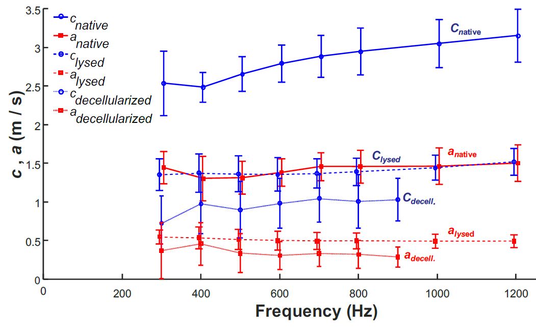
Livers were harvested from Lewis rats and animal protocols were approved by the State Office of Health and Local Affairs (LAGeSo, Berlin, Germany; Reg. No L 0421/12). Each rat liver sample was introduced into the cylindrical glass capillary and prepared for MRE. Native samples were prepared immediately after euthanization of the animal. Lysed samples from the same rat were embedded in PBS and stored overnight at -20℃ to destroy cells without removing cell debris. The samples were thawed at 4℃the next day and MRE measurements were ran at room temperature. Decellularized livers were prepared from additional rats, detailed procedures have been published elsewhere [3]. A compact MRE tabletop device with a 0.5 T permanent-magnet based MRI system was used for the MRE experiments. Details of the system are described in [4, 5]. MRE was performed in native, lysed and decellularized rat liver samples (width x height: 8 mm x 1 cm). The shear modulus dispersion functions were acquired at 300-1200 Hz. Two viscoelastic models were fitted to the data: i) the powerlaw springpot model (SP) comprising a shear modulus μSP and powerlaw exponent α and ii) the Kelvin-Voigt (KV) model comprising a shear modulus parameter μKV and a viscosity parameter η.Results:
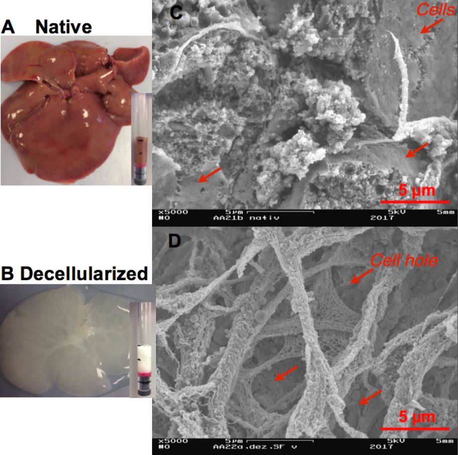
Decellularized tissue shows a rich collagen matrix with cavities due to the removed cells in SEM images (Figure 1). Cell lysis and complete cell removal drastically changed the mechanical properties of the liver. Overall, liver stiffness progressively decreased and the powerlaw coefficient α increased due to cell lysis or cell removal (Figure 2). The fit residue σ for the KV-model indicating the quality of the fit was lower in decellularized tissue than in lysed and native tissue (mean σ SP decellularized, lysed, native: 0.08 ± 0.04, 0.17 ± 0.04, 0.31 ± 0.03). Conversely, the SP-model similarly matched the viscoelastic properties of all three tissue states (mean σ SP decellularized, lysed, native: 0.11 ± 0.04, 0.12 ± 0.03, 0.11 ± 0.04).Discussion and conclusions:
Tabletop MRE can reproducibly measure the change of viscoelastic properties in liver tissue due to cell removal or modification of cellular integrity. Overall, decellularized tissue is much softer than native tissue and the degree of stiffness reduction is similar to what is obtained by freezing and thawing of the tissue. However, the parameter changes with frequency, i.e. the viscoelastic dispersion function was distinct in all three investigated tissue states. While the viscoelastic properties of intact liver tissue are determined by cells and can be described by a power law behavior, decellularized tissue has more solid-like properties which are better described by the KV-model. Our study contributes to research on the mechanical interactions between cells and extracellular matrix in liver tissue. Our results indicate an important contribution of cell membrane integrity to the global stiffness of non-fibrotic liver tissue and indicates that elastography could be sensitive to hepatic cell changes such as cell ballooning in non-alcoholic steatohepatitis (NASH). Furthermore, the presented results indicate the usefulness of MRE in regenerative tissue research.Acknowledgements
No acknowledgement found.References
[1] Badylak Stephen F,Taylor D and Uygun Korkut. Whole-Organ tissue engineering: Decellularization and recellularization of Three-dimensional matrix scaffolds. Annu. Rev. Biomed. Eng. 2011; 13 (27-53).
[2] Uygun B.E, Soto-Gutierrez A, Yagi H, et al. Organ reengineering through development of a transplantable recellularization liver graft using decellularized liver matrix. Nature medicine 2010; 16 (814-820).
[3] Struecker B, Butter A, Hillebrandt K, et al. Improved rat liver decellularization by arterial perfusion under oscillating pressure conditions. J. tissue engineering and regenerative medicine 2017; 11 (531-541).
[4] Ipek-Ugay S, Driessle T, Ledwig M, et al. Tabletop magnetic resonance elastography for the measurement of viscoelastic parameters of small tissue samples. J. Magnetic Resonance 2015; 251 (8-13).
[5] Braun J, Tzschätzsch H, Korting C, et al. A compact 0.5 T MR elastography device and its application for studying viscoelasticity changes in biological tissues during progressive formalin fixation. Magn Reson Med 2017.
Figures