5584
Ristretto MRE: A Generalized Multi-Shot GRE-MRE Sequence1Institute for Biomedical Engineering, University and ETH Zurich, Zurich, Switzerland, 2Division of Research Oncology, Guy's and St Thomas' NHS Foundation Trust, London, United Kingdom, 3Division of Imaging Sciences and Biomedical Engineering, King's College London, London, United Kingdom
Synopsis
The purpose of this work is to increase GRE-MRE sequence flexibility by generalizing the multi-shot eXpresso approach. We show that Ristretto MRE allows for the fine-tuning of imaging shot durations in both multi-slice and 3D-MRE acquisitions permitting significant scan time reductions without loss of image quality.
Introduction
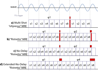
In multi-slice MRE1,$$$N_S$$$ slice excitations are interleaved to maximize the per-slice repetition time, while synchronization to the wave period$$T=f^{-1}$$needs to be ensured. In order to reduce total scan duration, the “eXpresso” sequence was proposed acquiring an integer number of $$$x$$$ slices per wave period2 as illustrated in Figure 1a. Different wave offsets are acquired for each k-line by introducing a delay $$$T/N_P$$$, which is depicted in red. Since each slice is acquired with a different phase shift relative to the onset of wave generation, phase correction is necessary before wave inversion. While the eXpresso scheme has been used in numerous MRE applications2–5, it lacks flexibility, which in turn may result in extended scan durations. The objective of the present work is to show that the eXpresso scheme is overly restrictive and to propose a generalized multi-shot approach that allows for the fine-tuning of image shot durations in multi-slice and 3D GRE-MRE acquisitions without compromising image quality.Theory
The
“Ristretto” concept generalizes the eXpresso approach by acquiring an arbitrary
number of slices $$$N_S$$$ within an arbitrary number of wave periods $$$N_W$$$
(Figure 1b). Hence, the number of slices per period is
allowed to be a rational number, which is given by$$x=N_S/N_W,$$while each imaging shot is of duration$$T_R=T/x.$$In
addition, the delay can be omitted by distributing it evenly over all imaging
shots (Figure 1c). By interleaving the wave phases through
extending the delay by an integer factor $$$N_D$$$ (Figure 1d), the flexibility of the imaging shot duration
can be further increased to $$T_R=T\left(\frac{N_W}{N_S}+\frac{N_D}{N_PN_S}\right).$$N.B.:
$$$N_D$$$ must be chosen such that all phase offsets are acquired. The allowed
$$$N_D$$$ are given by integers not divisible by any prime factor of $$$N_P$$$.
If $$$N_P$$$ is a power of 2, the delay $$$N_D$$$ can be any uneven number and
the repetition time can be chosen in steps of$$\Delta{}T_R=\frac{2T}{N_SN_P}.$$In a typical 10 slice, 8 phase acquisition, this amounts to
$$\Delta{}T_R^{N_S=10,N_P=8}=\frac{T}{40}.$$
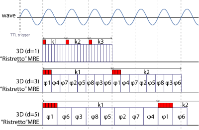
The extended-no-delay-Ristretto scheme can be applied to 3D
encoding (Figure
2),
where a slab is excited and phase encoding is employed along the slice
direction with interleaved acquisition of the motion phases for the same $$$k_y$$$/$$$k_z$$$-lines.
The shot duration is given by$$T_R=T\frac{N_D}{N_P},$$
with $$$N_W=N_D$$$, while the same rule applies for
$$$N_D$$$ as with multi-slice Ristretto.
Methods
The proposed Ristretto sequences were implemented on different Philips platforms and compared to eXpresso MRE in an ultrasound gel phantom, in the breast and the thigh of one volunteer. Sequence parameters in each of the three experiments were held constant, except for Ristretto parameters and slice TRs, respectively. Scan parameters and actuation modalities are included in the result figures for each scan. Shear wave velocities were obtained using a FEM-based inversion algorithm6.Results & Discussion
In Figure 3, the real-part of the complex displacement field of an ultrasound gel phantom is shown. eXpresso, Ristretto and no-delay-Ristretto show excellent agreement. With the chosen echo time of 9.2ms and 30 Hz wave frequency, eXpresso MRE only permits two shots per period. The use of Ristretto allows for 2.5 slices per period, reducing scan duration to 80%, while interleaved acquisition of the eight phases further reduces scan duration to 65%.
In Figure 4, fractional GRE-MRE is compared to multi-slice Ristretto and 3D Ristretto in the thigh. Displacement fields are in good agreement between all three techniques. eXpresso MRE cannot be applied as the high wave frequency does not allow for the acquisition of multiple slices per period. Multi-slice Ristretto can acquire 12 slices within 10.625 wave periods by interleaving the phase offsets. 3D Ristretto with $$$N_W=7$$$ acquires the eight phases in reverse ordering.
In Figure 5, the standard eXpresso breast protocol using 3 shots per period is compared to multi-slice Ristretto, 3D Ristretto and a rapid 3D Ristretto scheme. Displacement fields and reconstructed shear wave velocities are in good agreement within the breast, however, the increased SNR in the 3D scans allows for the reconstruction of the stiffer areas in the region of the axial lymph nodes. By reducing the number of phases to four, making use of the increased encoding efficiency of Hadamard encoding7, and the high SNR of 3D acquisitions, 4D MRE of the breast and axial lymph nodes becomes feasible in under 2 minutes.
Conclusions
We have demonstrated, that the multi-shot concept of eXpresso MRE can be extended to allow for flexible tuning of imaging shot durations and permits reduction of total scan time without limiting constraints on the number of shots, slices, or the specific choice of the echo time. The concept can be further extended to 3D GRE-MRE and in combination with Hadamard encoding and the acquisition of four wave phases, allows for 4D MRE of the breast in under 2 minutes.Acknowledgements
This project has received funding from the European Union’s Horizon 2020 research and innovation programme under grant agreement No 668039.References
1. Muthupillai R, Lomas D, Rossman P, Greenleaf J, Manduca A, Ehman R. Magnetic resonance elastography by direct visualization of propagating acoustic strain waves. Science (80- ). 1995;269(5232):1854-1857. doi:10.1126/science.7569924.
2. Garteiser P, Sahebjavaher RS, Ter Beek LC, et al. Rapid acquisition of multifrequency, multislice and multidirectional MR elastography data with a fractionally encoded gradient echo sequence. NMR Biomed. 2013;26(10):1326-1335. doi:10.1002/nbm.2958.
3. Ronot M, Lambert S, Elkrief L, et al. Assessment of portal hypertension and high-risk oesophageal varices with liver and spleen three-dimensional multifrequency MR elastography in liver cirrhosis. Eur Radiol. 2014;24(6):1394-1402. doi:10.1007/s00330-014-3124-y.
4. Hatt A, Cheng S, Tan K, Sinkus R, Bilston LE. MR Elastography Can Be Used to Measure Brain Stiffness Changes as a Result of Altered Cranial Venous Drainage During Jugular Compression. Am J Neuroradiol. 2015;36(10):1971-1977. doi:10.3174/ajnr.A4361.
5. Hoelzl S, Sethi S, Sudakova J, et al. Enabling High Resolution MRE Images of the Breast. In: Intl. Soc. Mag. Reson. Med. 25. Honolulu; 2017:4938.
6. Fovargue D, Sinkus R, Nordsletten D. Robust MR Elastography Stiffness Quantification Using a Localized Divergence Free Finite Element Reconstruction. In: Intl. Soc. Mag. Reson. Med. 25. Honolulu; 2017:1368.
7. Guenthner C, Runge JH, Sinkus R, Kozerke S. Hadamard Encoding for Magnetic Resonance Elastography. In: Intl. Soc. Mag. Reson. Med. 25. Honolulu; 2017:1378.
8. Runge J, Hoelzl S, Sudakova J, et al. A Novel MR Elastography Transducer Concept Based on a Rotational Eccentric Mass: The Gravitational Transducer. In: Intl. Soc. Mag. Reson. Med. 25. Vol 1369. Honolulu; 2017.
Figures

