5581
TURBINE-MRE: A 3D Hybrid Radial-Cartesian EPI Acquisition for MR Elastography1Radiology, Mayo Clinic, Rochester, MN, United States
Synopsis
A 3D gradient-echo EPI MR Elastography pulse sequence has been developed using a hybrid radial-Cartesian readout scheme, named Trajectory Using Radially Batched Internal Navigator Echoes (TURBINE). The feasibility of this TURBINE-MRE sequence was demonstrated in a phantom study.
Introduction
Spin-Echo EPI has been widely used for MR Elastography (MRE) acquisitions in the brain, liver, and heart (1). However, this method is susceptible to cardiac and respiratory motion. For these applications, multiple breath holds, which can be challenging for many patients, are typically required to produce artifact-free images. Recently, a 3D hybrid radial-Cartesian readout scheme, named Trajectory Using Radially Batched Internal Navigator Echoes (TURBINE), has been introduced for brain diffusion imaging (2) and fMRI (3). In a TURBINE acquisition, 2D EPI planes are rotated in k-space by different angles about the phase-encoding axis to provide a dataset that is radially sampled in two dimensions and Cartesian sampled in the other. When executed in a continuous manner using golden-angle plane rotations, this partial radial acquisition scheme is, in theory, self-navigated and, through a synergistic combination of retrospective respiratory gating and advanced reconstruction, enables free-breathing imaging(4). Such capabilities could greatly benefit both cardiac and abdominal MRE applications. In this study, we report on the implementation of a TURBINE-MRE acquisition scheme and demonstrate its feasibility in a phantom study.Methods
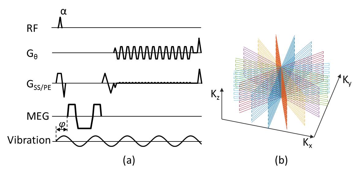
The proposed TURBINE-MRE acquisition strategy is based on the GRE-EPI MRE sequence shown in Fig. 1a. MRE motion-encoding gradients (MEGs) were added prior to the EPI readout to encode MRE harmonic tissue motion into the image phase, and they can be applied on the x, y, and/or z axes to record the full vector motion. MEGs and the mechanical vibration were temporally shifted by various amounts, φ, in different acquisitions to acquire wave images at different phases of the harmonic motion. The EPI readout axis, Gθ, rotated about the slab-selective (GSS)/phase-encoding (GPE) axes in successive TRs. One 2D plane within the target 3D k-space was sampled with the EPI trajectory (TURBINE blade) during each TR (Fig. 1b). To cover all of 3D k-space, the sampling plane was angularly rotated by a fixed increment before each repetition. Each EPI blade was first reconstructed using a standard Cartesian EPI reconstruction pipeline (5,6). Reconstruction of the radially sampled plane (orthogonal to the phase-encoding axis) was then performed via density-compensated gridding using the non-uniform FFT (NUFFT)(7,8). Finally, the MRE phase signal was jointly estimated from the set of reconstructed complex coil images via maximum likelihood (ML) estimation (9). A PVC phantom was scanned on a 1.5T GE scanner with an 8-channel head coil. External vibration at 140 Hz was applied in the y-direction. The imaging parameters were: TR/TE=50/18.5 ms, flip angle=40°, EPI FOV = 260 (read) x 100 (phase) mm2, acquisition matrix = 80×32. 128 radial-encoding positions were evenly distributed over 180 degrees. The reconstructed image spatial resolution was an isotropic 3.25 mm. The total scan time per 3D volume was 6.4 seconds, or 2:34 minutes to obtain a complete MRE dataset with 6 motion-encoding directions (±X, ±Y, and ±Z) and 4 time offsets (24 3D volumes total). For comparison, a standard 2D, multislice, spin-echo (SE) EPI-MRE acquisition was also performed with the same imaging parameters.Results
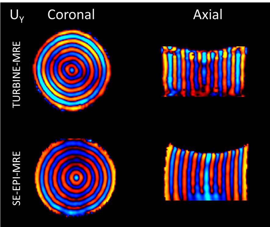
Figure 2 shows the first harmonic of the 3D wave images obtained from the TURBINE-MRE and SE-EPI-MRE phase images. Coronal and axial views are shown. Of note, TURBINE-MRE is a true 3D acquisition, whereas SE-EPI-MRE is a multislice acquisition which produces a stack of 2D images.
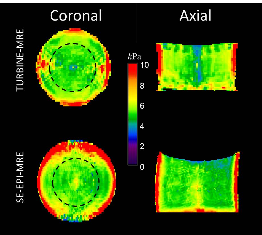
Figure 3 shows the stiffness maps calculated using a local-frequency estimation algorithm (LFE) (10) for the two sequences. The mean stiffness from TURBINE-MRE and SE-EPI-MRE in the ROI (black circle) was 5.2±0.5 kPa and 5.1±0.4 kPa, respectively.
Discussion and Conclusion
This phantom study demonstrates the feasibility of TURBINE-MRE for acquiring true 3D wave information in a highly efficient manner, and provides results that are consistent with those of the standard SE-EPI-MRE sequence (<2% difference). This sequence will particularly help the cardiac MRE (cMRE), which, when using 2D EPI-based acquisitions, faces additional challenges due to the simultaneous presence of cardiac and respiratory motion. A typical cMRE sequence acquires only one cardiac phase within a breath-hold (11). The proposed TURBINE-MRE sequence provides a foundation upon which a free-breathing, cine-type MRE acquisition could be built, using a continuous golden-angle rotation scheme, retrospective cardiac and respiratory binning of k-space data in discrete motion states, and compressed sensing reconstruction (4).Acknowledgements
This work was supported by National Institutes of Health (NIH) grants 5R01HL115144 and EB001981 and Mayo Clinic Department of Radiology internal funding.References
- Glaser KJ, Manduca A, Ehman RL. Review of MR elastography applications and recent developments. J Magn Reson Imaging 2012;36(4):757-774.
- McNab JA, Gallichan D, Miller KL. 3D steady-state diffusion-weighted imaging with trajectory using radially batched internal navigator echoes (TURBINE). Magn Reson Med 2010;63(1):235-242.
- Graedel NN, McNab JA, Chiew M, Miller KL. Motion correction for functional MRI with three-dimensional hybrid radial-Cartesian EPI. Magn Reson Med 2016.
- Feng L, Axel L, Chandarana H, Block KT, Sodickson DK, Otazo R. XD-GRASP: Golden-angle radial MRI with reconstruction of extra motion-state dimensions using compressed sensing. Magn Reson Med 2016;75(2):775-788.
- Bruder H, Fischer H, Reinfelder HE, Schmitt F. Image reconstruction for echo planar imaging with nonequidistant k-space sampling. Magn Reson Med 1992;23(2):311-323.
- Hinks RS, Mock BJ, Frigo FJ, Zhao X; Method and apparatus of echo planar imaging with real-time determination of phase correction coefficients 2007.
- Pipe JG, Menon P. Sampling density compensation in MRI: Rationale and an iterative numerical solution. Magnetic Resonance in Medicine 1999;41(1):179-186.
- Fessler JA, Sutton BP. Nonuniform fast Fourier transforms using min-max interpolation. Ieee T Signal Proces 2003;51(2):560-574.
- Bernstein MA, Grgic M, Brosnan TJ, Pelc NJ. Reconstructions of phase contrast, phased array multicoil data. Magn Reson Med 1994;32(3):330-334.
- Manduca A, Oliphant TE, Dresner MA, Mahowald JL, Kruse SA, Amromin E, Felmlee JP, Greenleaf JF, Ehman RL. Magnetic resonance elastography: Non-invasive mapping of tissue elasticity. Medical Image Analysis 2001;5(4):237-254.
- Arani A, Glaser KL, Arunachalam SP, Rossman PJ, Lake DS, Trzasko JD, Manduca A, McGee KP, Ehman RL, Araoz PA. In vivo, high-frequency three-dimensional cardiac MR elastography: Feasibility in normal volunteers. Magn Reson Med 2016.
Figures