5573
Alterations of regional homogeneity in poststroke aphasia1Shanghai Key Laboratory of Magnetic Resonance and Department of Physics, School of Physics and Materials Science, East China Normal University, Shanghai, China, 2Department of Rehabilitation Medicine, Huashan Hospital, Shanghai, China
Synopsis
The purpose of this study was to investigate intrinsic local synchrony changes in poststroke aphasia patients during resting-state fMRI scans. Fifteen patients (aged, 39-62 years, 4 female) and 30 age- and gender-matched healthy controls participated. Regional homogeneity (ReHo) was calculated to measure spontaneous brain activity. The results showed that poststroke aphasia patients exhibited significantly increased ReHo in the left frontal lobe, left cingulate gyrus, left corpus callosum, left temporo-parietal areas and right middle frontal gurus. Our study showed patterns of intrinsic local synchronization are altered in poststroke aphasia patients at resting state.
Purpose
Poststroke aphasia is a significant clinical problem that is usually caused by left hemisphere lesions. Numerous functional neuroimaging are available for the investigation of the language architecture and the neurobiological mechanism underlying poststroke aphasia. However, information regarding local synchronization of spontaneous functional magnetic resonance imaging blood–oxygen level-dependent fluctuations in poststroke aphasia was limited. The aim of this study was to investigate the regional homogeneity (ReHo) in poststroke aphasia patients under resting-state.Materials and Methods
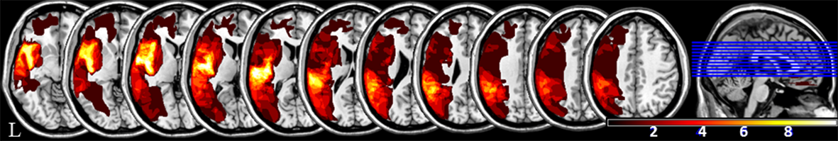
Fifteen patients with poststroke aphasia (aged, 39-62 years, 4 female) and 30 age- and gender-matched healthy controls participated in this study. All patients were first stroke and the lesion was located in the left hemisphere, and they had no movement function and other obstacles. Functional images were acquired using an EPI sequence with the following parameters: TR/ TE = 2000/ 30 ms, 210 volumes. Structural scans included a high-resolution three-dimensional T1-weighted magnetization-prepared rapid-acquisition gradient-echo pulse sequence (TR/ TE = 2530/ 2.34 ms, 192 slices). The doctor manually traced the outline of the lesion on individual 3D T1 images using MRIcron, thereby creating a lesion mask for each patient. After the spatial normalization process, the union of all individual lesion masks was used to construct a group lesion mask for the patients (Figure1). We calculated ReHo (0.01~0.08 Hz) to measure spontaneous brain activity using Data Processing Assistant for Resting-State fMRI software. The maps of the significant differences in ReHo of the 15 aphasic patients and the 30 controls were compared using voxel-wise two-sample t tests, and the group comparison was applied within the patients’ group masks to exclude the lesions in all patients.Results
When compared to healthy controls, poststroke aphasia patients exhibited significantly increased ReHo in the left frontal lobe, left cingulate gyrus, left corpus callosum, left temporo-parietal areas and right middle frontal gyrus.Discussion
Aphasic patients exhibited significantly increased local synchronization in the left cingulate gyrus, left corpus callosum, left temporo-parietal areas than healthy controls, these areas all around lesion areas. The ReHo value also increased in the right middle frontal gyrus, which is homologous to left-hemisphere language network.1,2 The results suggest that aphasic patients’ adaptive changes were associated with alterations of spontaneous brain activity in the left hemisphere area around the lesion and right hemisphere areas homologous to left-hemisphere language region during chronic phase.Acknowledgements
This research was supported by grants from the National Natural Science Foundation of China (NO. 81571658 to X. X. Du)References
1. Klingbeil J, Wawrzyniak M, Stockert A, Saur D (2017) Resting-state functional connectivity: An emerging method for the study of language networks in post-stroke aphasia. Brain and cognition. DOI:10.1016/j.bandc.2017.08.005
2. Turken AU, Dronkers NF (2011) The neural architecture of the language comprehension network: converging evidence from lesion and connectivity analyses. Frontiers in systems neuroscience 5:1.
Figures