5569
Novel MRI-compatible foot-sole stimulator for characterizing the cortical regions pertaining to walking-related somatosensory perception1Academy for Advanced Interdisciplinary Studies, Peking University, Beijing, China, 2College of Engineering, Peking University, Beijing, China, 3Department of Radiology, Peking University First Hospital, Beijing, China, 4Hebrew SeniorLife Institute for Aging Research, Roslindale, MA, United States, 5Division of Gerontology, Beth Israel Deaconess Medical Center, Boston, MA, United States, 6Harvard Medical School, Boston, MA, United States
Synopsis
The decline of foot-sole somatosensation is one of the main contributors to diminished balance of walking. The response of brain regions to the inputs from foot-soles is critical to the somatosensory perception, which, however, remains unknown. Here we developed a dual-drive stimulator applying pressure stimuli that mimic those as experienced during walking on the feet. This stimulator is compatible for MRI and the foot-sole pressure stimuli significantly increases the excitability (i.e., BOLD signal intensity) within multiple brain regions. This stimulator may thus be a novel tool to characterize the brain’s responsiveness in the perception of foot-sole somatosensation pertaining to walking.
Introduction
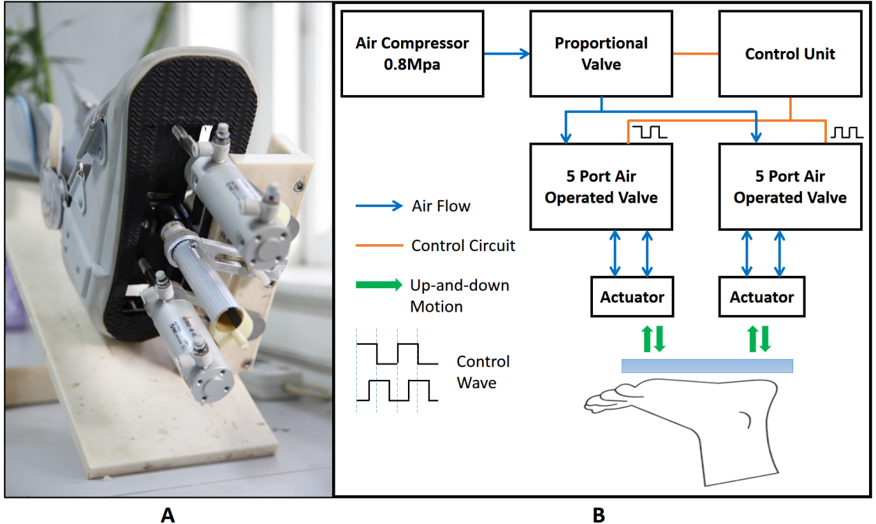
The decline in foot-sole somatosensation is highly prevalent in aging and diseases and is one of the main contributors to the diminished balance and increased risk of falls in older adults [1]. When walking on the ground, somatosensory inputs arise from foot soles and activate spinal and supraspinal networks. The responsiveness of brain regions to these afferent inputs is thus important for the somatosensory perception, which, however, remains not fully understood. Hao et al., recently developed a foot-sole pressure stimulation system applying programmable single-point stimuli on the front foot soles and observed that several cortical regions were activated by the applied stimulus, as measured by the intensity of blood oxygenation level-dependent signal (BOLD) [2]. Here we developed a novel foot-sole pressure stimulator (FIG.1), which is able to program the pressure to those as experienced by foot soles during walking on the ground, and investigated its compatibility in MRI scanning environment and the excitability of cortical regions when perceiving the pressure stimulus.Methods
This stimulator (FIG.1) was equipped with dual pneumatic actuators. One control unit placed five meters away was attached to the aluminum pneumatic actuators and plastic platform via plastic air tubes, and regulated the stimulator to apply the programmable pressure in one-degree-of-freedom oscillations on the front sole and heel of foot soles.
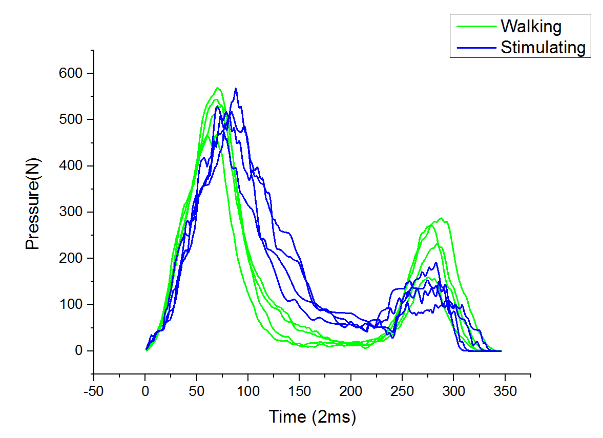
1) To examine the similarity between the programmed pressure stimulus and the pressure experienced during actual walking, five healthy younger adults completed four 5-meter straight walking trials at their preferred walking speed. The pressure on their foot soles was measured using pressure insoles. We then programmed the pressure stimulus based upon the measured pressure and applied it on the foot soles when the participants were laying as during the MRI scan. The pressure stimulus on the foot soles were measured by the same insoles and then compared to the “real” pressure.
2) To test the MRI compatibility of this stimulator, we obtained five sets of phantom images in each of the three following conditions: no stimulator was in MRI scanner room, stimulator was working and off in the scanner room.
3) To explore the cortical response to the foot-sole stimulation, nine healthy younger adults completed 3 3.5-minute block-designed sequences of fMRI. Each sequence contained alternating 30-second blocks of foot stimulation and no foot stimulation. Before the MRI test, the pressure during walking was measured for each individual. During foot stimulation blocks, the foot-sole stimulator applied the programmed pressure, which mimics the pressure measured during walking, to the right foot sole. The BOLD signals in each block were recorded.
Results
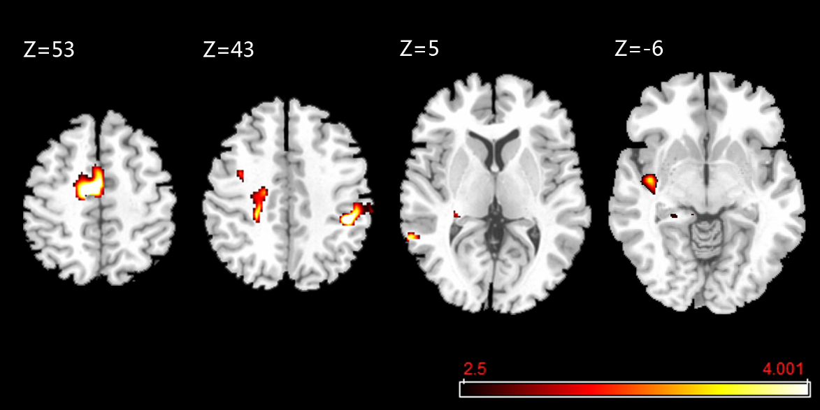
We observed that: 1) the programmed pressure correlated with the actual pressure during walking (mean r=0.75±0.14, P<0.01) (FIG.2); 2) no significance of signal to noise ratio in the phantom images between situations (F=0.32, P=0.72); and 3) compared to rest, the excitability of supplementary motor area of left medial frontal gyrus, supramarginal gyrus of right inferior parietal lobule, paracingulate gyri, left insula, precentral gyrus, middle temporal gyrus and hippocampus of left parahippocampal gyrus were significantly increased (uncorrected P<0.001, k≥10)(FIG.3).Discussion
This piloting study has demonstrated that the novel dual-drive foot sole stimulator can simulate the oscillations of pressure on foot soles as those experienced during walking on the ground, and has no impact on the image quality when it is working during the MRI scan. Besides the previously identified brain regions involving in the perception of somatosensation, we have observed several other regions are activated when particularly perceiving this walking-related foot sole stimulation, such as supplementary motor area, precentral gyrus, etc.. This indicates that these regions may uniquely contribute to the neurophysiologic control of walking, which is warranted to be explored in future studies.Conclusion
Our novel dual-drive foot sole stimulator enables characterizing the brain’s responsiveness to those important somatosensory inputs, and therefore, may serve as a novel tool to study the characteristics of functional brain networks involved in the perception and modulation of walking-related foot-sole pressure in those vulnerary populations, such as people with diabetic peripheral neuropathy.Acknowledgements
No acknowledgement found.References
[1] Manor B, Costa M D, Hu K, et al. Physiological complexity and system adaptability: evidence from postural control dynamics of older adults[J]. Journal of Applied Physiology, 2010, 109(6): 1786-1791.
[2] Hao Y, Manor B, Liu J, et al. Novel MRI‐compatible tactile stimulator for cortical mapping of foot sole pressure stimuli with fMRI[J]. Magnetic resonance in medicine, 2013, 69(4): 1194-1199.
Figures