5560
Dynamic development of spontaneous neuronal activity in postpartum women: A longitudinal resting-state functional MRI study1Department of Electronic Science, Xiamen University, Xiamen, China, 2Shanghai Key Laboratory of Magnetic Resonance and Department of Physics, School of Physics and Materials Science, East China Normal University, Shanghai, China
Synopsis
Investigating the neuroanatomical and functional bases in postpartum women have contributed to understanding the processing of cognitive and emotional alterations. The objective of this study was to examine spontaneous neuronal activity and functional connectivity of resting-state networks in postpartum women using a longitudinal resting-state functional magnetic resonance imaging. Twenty-three postpartum women were recruited.
Introduction
Women will undergo numerous changes including behaviours, emotions, physical, hormone, and among others during the pregnant and postpartum periods. We previously demonstrated that in the late postpartum women, brain structural plasticity exists compared to the early postpartum women using a longitudinal voxel-based morphometric study. In the current study, we look at possible changes in spontaneous neuronal activity including the amplitude of low-frequency fluctuation (ALFF), regional homogeneity (ReHo) and functional connectivity occurring longitudinally over a period of two years. Second, we aim to determine the relationships of spontaneous neuronal activity and empathetic abilities in postpartum women.Materials and Methods
Twenty-three postpartum women (mean age: 30.2 ± 2.0 years) were selected for this longitudinal functional magnetic resonance imaging (fMRI) study. All subjects were scanned twice: the first scan (scan 1) was collected one year after delivery, and the second scan (scan 2) was performed two years after delivery. The average time interval between scans was 704 days (SD = 2.0 days). MRI scanning sessions were performed on a Siemens 3.0 T Trio Tim MR system using a 12-channel head coil with the following acquisitions: a high-resolution three-dimensional T1-weighted sequence images were acquired for anatomical reference (TR = 2530 ms, TE = 2.34 ms, 192 slices) and resting-state T2∗-weighted images with the following parameters TR = 2000ms, TE = 30 ms, 33 slices, 210 volumes were acquired. Subjects were instructed to relax and rest, with their eyes closed during the resting-state fMRI scan. ALFF (0.01 - 0.08 Hz) and ReHo were analyzed to measure spontaneous brain activity and functional connectivity was analyzed to investigate empathy-related brain network using Data Processing Analysis for Brain Imaging and Statistical Parametric Mapping software. We further investigated relationships between empathetic abilities and the differential brain regions showing altered ALFF in postpartum women.Results
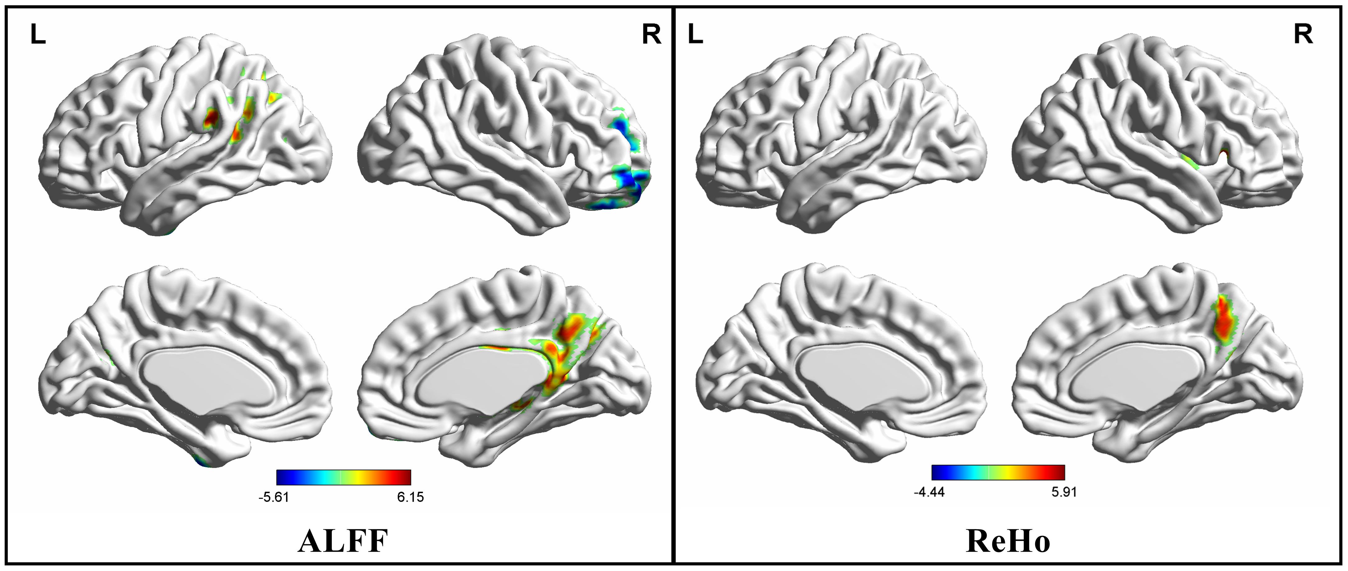
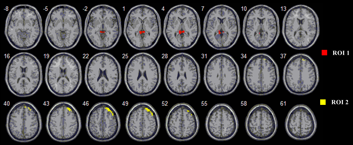
The results showed that ALFF increased in the superior medial prefrontal cortex and decreased in the precuneus, angular gyrus, supramarginal gyrus, hippocampus, posterior cingulate gyrus, thalamus, postcentral gyrus and inferior parietal lobule from scan 1 to scan 2. Decreased ReHo was found in the insula, inferior frontal gyrus, precentral gyrus, precuneus and posterior cingulate gyrus while increased ReHo was discovered in the left cerebellum posterior lobe (Figure 1). When significantly altered with ALFF of brain regions (e.g. precuneus, posterior cingulate gyrus) as the region of interest, postpartum women showed longitudinal altered connectivity regions in the frontal lobe and parietal lobe (e.g. superior middle frontal gyri, parahippocampal gyrus) (Figure 2). Correlation analysis showed significant correlations between the alterations of spontaneous neuronal activity and empathetic abilities in postpartum women.Discussion
Results demonstrated that spontaneous neuronal activity and functional connectivity altered in postpartum women from 1 to 2 years during nurturing infants. We reported altered ALFF and ReHo values in these regions such as supramarginal gyrus, angular gyrus and among others, all of which are related to higher-level cognitive functions and emotional processing1, 2. Functional connectivity analysis showed regions that were observed in ALFF analysis work in concert with other regions as a network that correlates with empathy. Our study first provides evidence for alterations spontaneous neuronal activity and brain network in postpartum women, which can implicate the neurophysiological mechanism in women after delivery. The detected changes suggested that postpartum women can exhibit functional reorganization, which is the dynamic development for adaptive changes including cognitive function and empathic processing over nurturing infants.Acknowledgements
This work was supported by the National Natural Science Foundation of China (grants 81571658 and U1632274) and the Social Science Foundation of China (grant 15ZDB016).References
1. Seghier ML. The angular gyrus: Multiple functions and multiple subdivisions. neuroscientist 2013; 19(1): 43-61.
2. Silani G, et al. Right supramarginal gyrus is crucial to overcome emotional egocentricity bias in social judgments. J Neurosci 2013; 33(39): 15466-76.
Figures