5553
Enhanced task-related frontoparietal functional connectivity during a n-back task in children with abacus training1Department of Physics, Zhejiang University, Hangzhou, China, 2Department of Optical Engineering, Zhejiang University, Hangzhou, China
Synopsis
Abacus-based mental calculation (AMC) training has the potential to induce cognitive plasticity. However, the neural correlates of such benefits remain unknown. In this study, we aimed to examine impacts of AMC training on task-related functional connectivity. We obtained functional imaging data from 23 children with AMC training and 21 control children. For AMC children, we found a increase in task-related functional connectivity within the frontoparietal regions, which was also significantly correlated with the speed in the 2-back condition. Therefore, AMC training may enhance the functional integration of frontoparietal circuity, and consequently result in improved task performance.
Introduction
There is a growing number of studies investigating plasticity induced by training at the behavioral level, as well as at the neural level[1]. Recently, abacus-based mental calculation (AMC), a method to perform fast and accurate arithmetic by manipulating beads on a imaginary abacus, has attracted intensive attention in the field of cognitive plasticity[2,3]. Here, we aimed to examine changes in task-related functional connectivity among children with AMC training, which may provide relevant information about the neural mechanisms underlying behavioral benefits.Method
Twenty-three children who received two hours per week of AMC training for about five years, and twenty-one control children who did not have any experiences of AMC participated in this study. Age, gender distributions, and intelligence did not differ significantly between the two groups. Imaging data were obtained using a 1.5T Philips MRI scanner. Whole brain T2*-weighted echo-planner images (EPI) were acquired for both the n-back task session: TR/TE = 2000/50 ms, flip angle = 90°, matrix size = 64 × 64, FOV = 230 mm × 230 mm, slice thickness/gap = 5 mm/0.8 mm, and slice numbers = 22). The n-back task consisted of four blocks (two 0-back and two 2-back). A high-resolution T1-weighted structural image was also obtained for each participant (TR/TE = 2500/4.6 ms, flip angle = 15°, matrix size = 256 × 256, FOV = 256 mm× 256 mm, voxel size = 1 × 1 × 1 mm3, and slice numbers = 150).Result
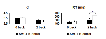
1. In the n-back task (Figure 1), a 2 (Group: AMC or control) × 2 (Condition: 0-back or 2-back) repeated measures ANOVA on discrimination index d’ (z target hit rate - z false alarm rate) yielded a significant main effect for Condition (F (1, 42) = 17.79, p < 0.001). The main effect for Group and the Group × Condition interaction did not reach significant. A parallel repeated measures ANOVA on RT revealed significant main effects for Group (F (1, 42) = 5.98, p < 0.05) and Condition (F (1, 42) = 22.45, p < 0.001). An interaction between Group and Condition was also present (F (1, 42) = 4.14, p < 0.05). Further analyses revealed shorter RTs in the AMC group than the control group in the 2-back condition (p < 0.05), but not in the 0-back condition (p = 0.13).
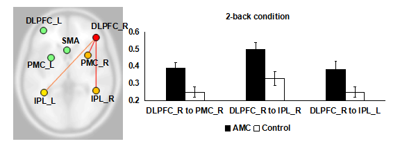
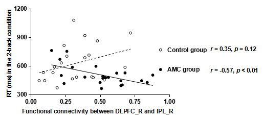
2. Seven regions of interest (ROIs, spheres of 8mm radius) were used as seeds to compare group differences in functional connectivity, which was implemented using the CONN toolbox[4]. These ROIs were centered on peak coordinates from a previous meta-analysis study[5] on n-back task-related activations, which also largely overlaped with task-related activations in the current study. For the 0-back condition, there were no significant group differences between any ROI-to-ROI functional connectivity. In the 2-back condition, compared to the controls, the AMC group showed greater functional connectivity between the right dorsolateral prefrontal cortex (DLPFC) and three other regions: right premotor cortex (PMC, t (42) = 2.90, p FDR corrected < 0.05), right inferior parietal lobule (IPL, t (42) = 3.03, p FDR corrected < 0.05), and left IPL (t (42) = 2.36, p FDR corrected < 0.05, Figure 2). In addition, for the AMC group, functional connectivity between the right DLPFC and the right IPL was significantly correlated with their RT in the 2-back condition (r = -0.57, p < 0.01, Figure 3). There were no significant performance-related functional connectivity for the control group.
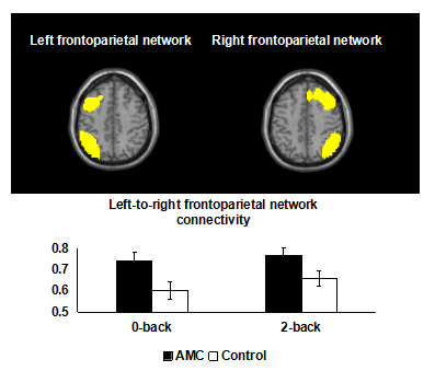
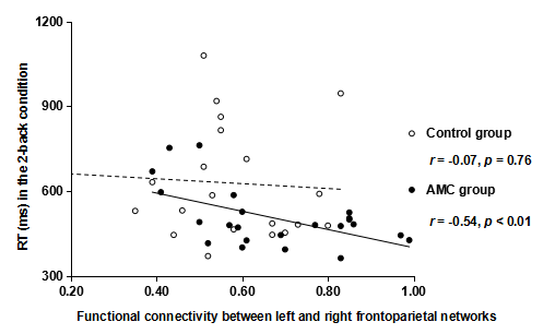
3. We also used independent component analysis to extract left and right frontoparietal networks. The two network maps were specified as ROIs to examine group differences in frontoparietal functional network interactions. In both 0-back and 2-back conditions, the AMC group showed increased functional connectivity of the left and right frontoparietal networks (Figure 4). For the AMC group, functional connectivity of the left and right frontoparietal networks in the 2-back condition was significantly correlated with their RT in the 2-back condition (Figure 5).
Discussion
Here we report novel results demonstrating training-related changes in frontoparietal functional connectivity during a n-back task. Given that the operation of AMC activates the frontoparietal areas largely overlapping with the working memory network[6], a possible explanation is that children with AMC training can utilize neural resources in the frontoparietal regions more cooperatively, which benefits task performance. These findings may have implications for helping individuals with cognitive deficits.Acknowledgements
The authors are very grateful to the Chinese Abacus and Mental Arithmetic Association, the Heilongjiang Abacus Association for their kind support. Thanks also to the children, parents, and teachers of Qiqihar for their participation in the study.References
1. Jolles D D, van Buchem M A, Crone E A, et al. Functional brain connectivity at rest changes after working memory training[J]. Human brain mapping, 2013, 34(2): 396-406.
2. Wang C, Geng F, Yao Y, et al. Abacus Training Affects Math and Task Switching Abilities and Modulates Their Relationships in Chinese Children[J]. PloS one, 2015, 10(10): e0139930.
3. Wang C, Weng J, Yao Y, et al. Effect of abacus training on executive function development and underlying neural correlates in Chinese children[J]. Human Brain Mapping, 2017, 38(10): 5234-5249.
4. Whitfield-Gabrieli S, Nieto-Castanon A. Conn: a functional connectivity toolbox for correlated and anticorrelated brain networks[J]. Brain connectivity, 2012, 2(3): 125-141.
5. Owen A M, McMillan K M, Laird A R, et al. N‐back working memory paradigm: A meta‐analysis of normative functional neuroimaging studies[J]. Human brain mapping, 2005, 25(1): 46-59.
6. Hanakawa T, Honda M, Okada T, et al. Neural correlates underlying mental calculation in abacus experts: a functional magnetic resonance imaging study[J]. Neuroimage, 2003, 19(2): 296-307.
Figures