5549
Altered structural covariance corticothalamic networks in epilepsy.1Depart. of Radiology, Jinling hospital, Nanjing, China, 2College Of Automation Engineering, Nanjing University of Aeronautics and Astronautics, Nanjing, China
Synopsis
It is the first work to depict the corticothalamus relationship by using the structural covariance connectivity. The epilepsy would significantly change the cortex-thalamus mode. The unilateral foci would lead the damage to the ipsilateral thalamus firstly. It provided a novel window to detected the mechanism of epilepsy.
Introduction
Patients with epilepsy can cause their brain functional and micro-structural damage. Thalamus as the key node in brain information exchange, plays an important role in the propagation of epileptic activity. Previous studies have confirmed that in various types of epilepsy, the structure and function of thalamus showed widely abnormal. Our group also found abnormal functional corticothalamic connectivity in patients with idiopathic generalized epilepsy (IGE). The structural covariance connectivity could describe the network pattern of brain micro-structural changes. Our previous research detected that the spread pattern of mesial temporal lobe epilepsy (mTLE) was from ipsilateral hippocampus to thalamus, finally to the frontal lobe. The functional corticothalamic connectivity has been used in the research of epilepsy, but the structural covariance connectivity has not been researched. Therefore, this work intended to use the structural covariance connectivity method to explore the pattern of corticothalamic microstructural changes, and its tendency in epilepsy.Material and Methods
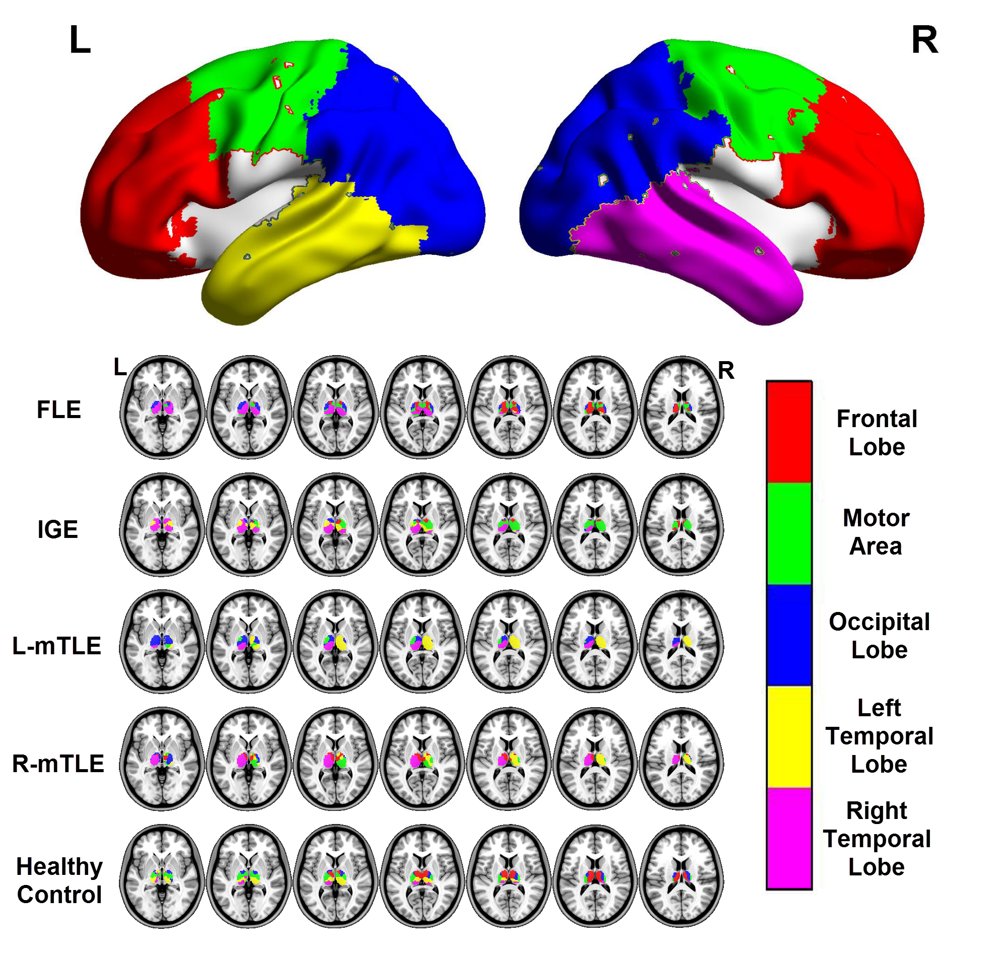
In this work, 392 patients with epilepsy (111 frontal lobe epilepsy, FLE, 111 IGE, 72 left mTLE and 68 right mTLE), and 111 healthy controls were included. The patients were diagnosed by neurologist in Jinling hospital. High resolution 3D-T1 weighted MRI was acquired at 3.0 T MRI scanner (SIEMENS Trio Tim, Germany). CAT12(http://www.neuro.uni-jena.de/cat/) was used to gained the gray matter maps. Firstly, the cortical cortex was separated into 5 ROIs according to the previous studies1. Then we combined the two motor ROIs and separated the temporal ROI into left and right ROIs.The thalamus was separated from AAL template. A winner-take-all strategy was used to divided the thalamus into 5 sub-regions according the highest correlation coefficient. The whole brain volume was removed during computing. These steps were done by our sorfware Across-Subject Brain Connect Analysis Toolbox (http://www.jlradiology.com/File.do).
Results
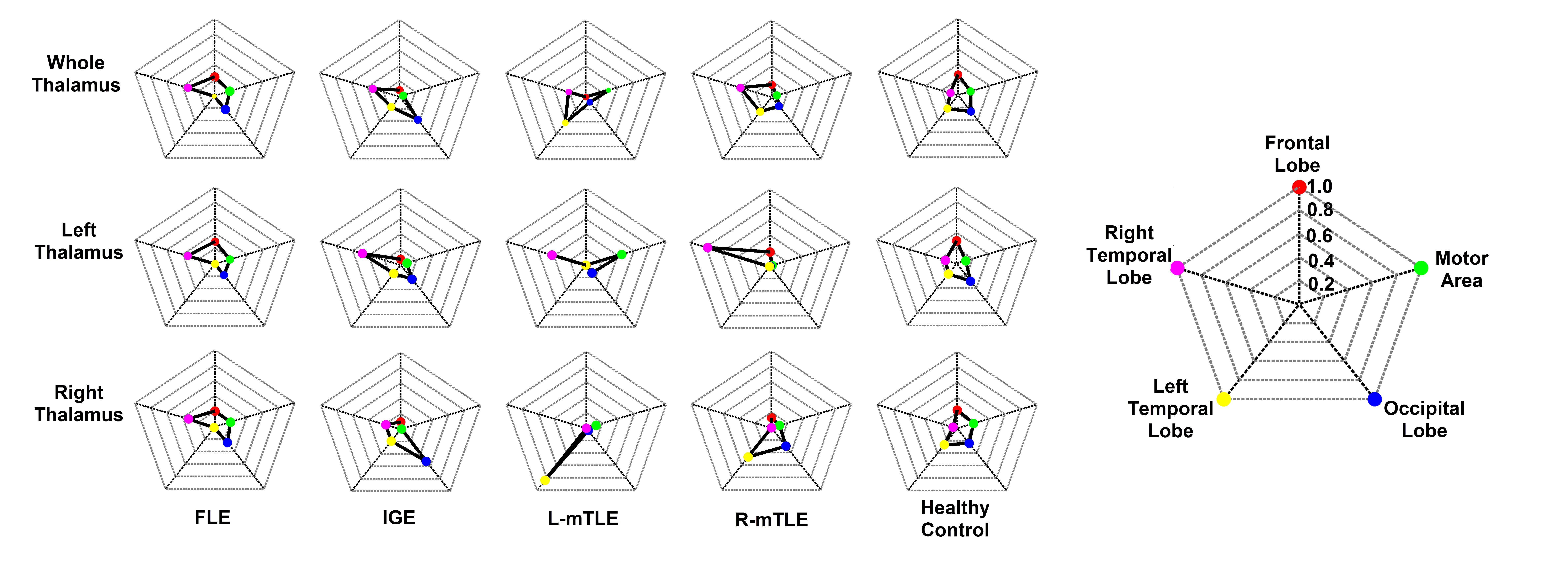
The corticothalamic structural covariance connectivity of healthy control (HC) showed that the thalamus could be separated into different regions based on the cortical ROIs. Compared to HC, FLE showed decreased connectivity between left temporal lobe and thalamus, and increased in right temporal lobe. The IGE group, L-mTLE group and R-mTLE group revealed lost connectivity between frontal lobe and thalamus. The connectivity numbers between motor area and thalamus in L-mTLE group increased significantly. The connectivity between right temporal lobe and thalamus was enhanced in all four types of epilepsy. After divided the thalamus into left and right ROIs, significant changes were found in L-mTLE and R-mTLE group. The connectivity from unilateral temporal to contralateral thalamus was disappeared.Conclusion
It is the first work to discusses the corticothalamus connectivity pattern based on the structural covariance connectivity. The epilepsy would significantly change the connectivity mode between cortex and thalamus. The results of L-mTLE and R-mTLE demonstrated that the unilateral epileptogenic foci would lead the damage to the ipsilateral thalamus firstly.Acknowledgements
This research was supported by the Natural Science Foundation of China (grants 81401402, Qiang Xu; 81401400, Gong-Jun Ji; 81471653, Wei Liao; 81422022, ZhiqiangZhang)References
1. Ji G J, Zhang Z, Xu Q, et al. Identifying corticothalamic network epicenters in patients with idiopathic generalized epilepsy[J]. American Journal of Neuroradiology, 2015, 36(8): 1494-1500.
2. Zhang Z, Liao W, Xu Q, et al. Hippocampus‐associated causal network of structural covariance measuring structural damage progression in temporal lobe epilepsy[J]. Human brain mapping, 2017, 38(2): 753-766.
Figures