5548
Regularized-Ncut: Robust functional parcellation of brain networks1Department of Radiology, Children's Hospital of Philadelphia, Philadelphia, PA, United States, 2Department of Radiology, Perelman School of Medicine, University of Pennsylvania, Philadelphia, PA, United States, 3Department of Bioengineering, School of Engineering and Applied Science, University of Pennsylvania, Philadelphia, PA, United States, 4Department of Biomedical Engineering, School of Medicine, Tsinghua University, Beijing, China
Synopsis
Human brain functional networks are critical in understanding intrinsic functional organization and systems. However, functional brain parcellation is affected by noise, resulting in artificial small patches and decreased functional homogeneity within certain networks. Using resting-state fMRI, we proposed a novel data-driven regularized-Ncut (RNcut) method by integrating a smoothing term and a small patches removal term to conventional Ncut for parcellating functional networks. The proposed method could delineate parcellated functional networks with higher functional homogeneity and better spatial contiguity with less noisy patches. A broad range of brain network applications and analyses could benefit from the proposed
Target audience
Neuroradiologists and neurologists.Purpose
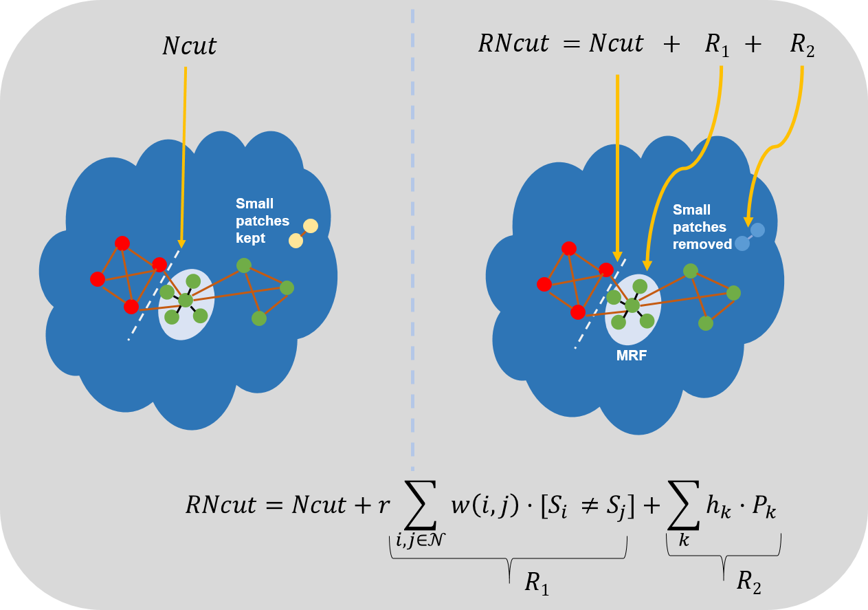
Human brain functional networks are critical in understanding intrinsic functional organization and systems. A variety of methods (e.g. [1-2]) have been developed to parcellate the brain into functional networks using resting-state fMRI (rs-fMRI). However, functional brain parcellation is affected by noise, resulting in small, artificial patches or networks with decreased functional homogeneity. In this study, we aimed to develop a novel data-driven Regularized-Ncut (RNcut) method by integrating a smoothing term and a small patches removal term into the conventional Ncut algorithm for parcellating functional networks. The experimental results demonstrated that the proposed method was more robust to noise and could generate the functional networks with higher functional homogeneity and spatial contiguity than competing methods.Methods
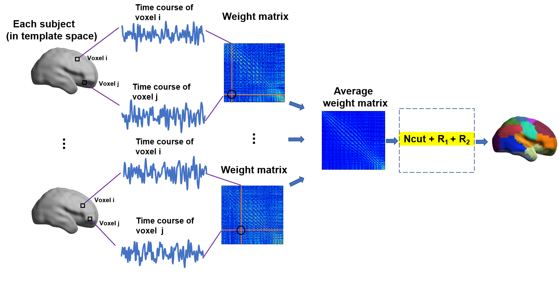
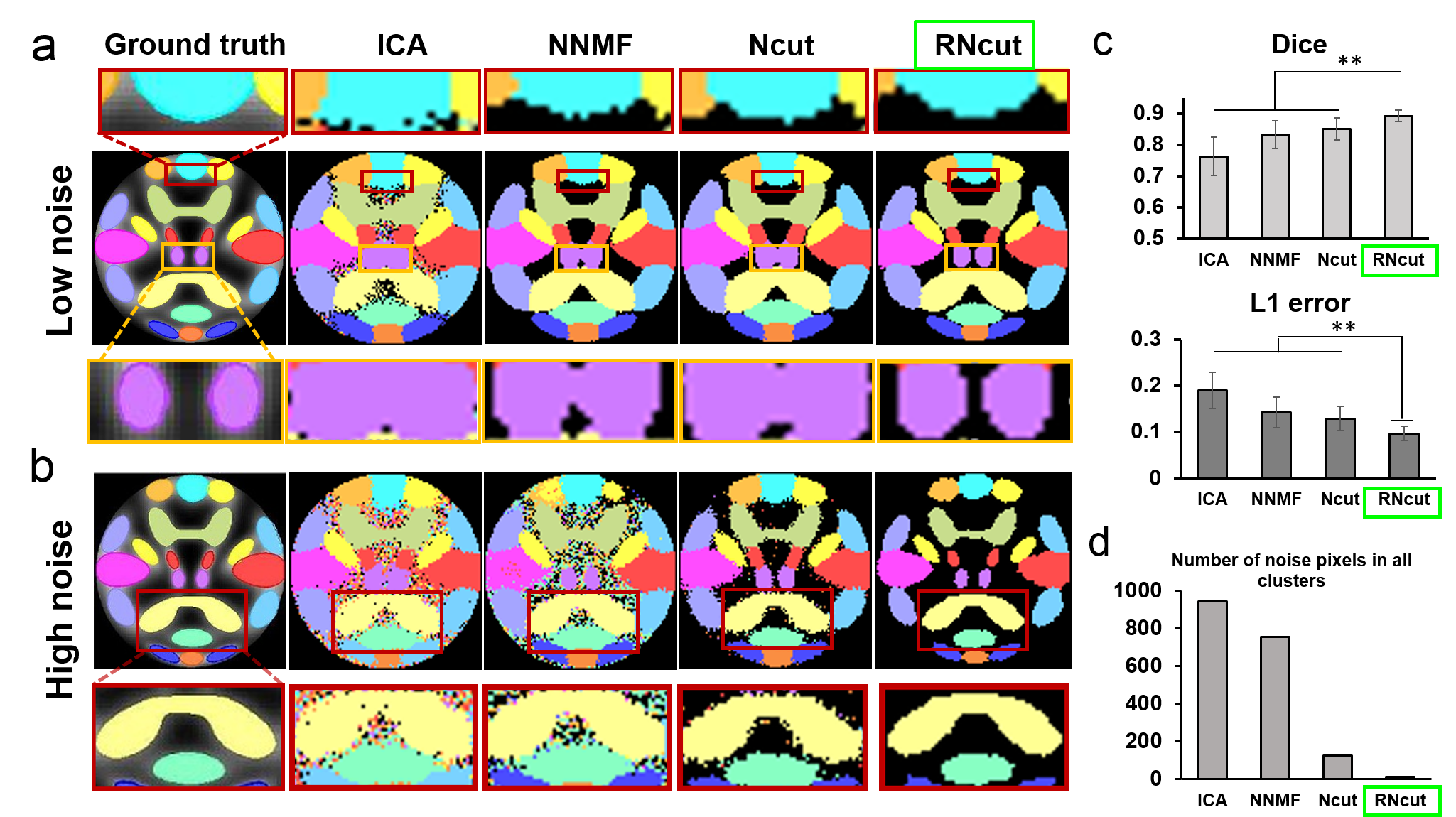
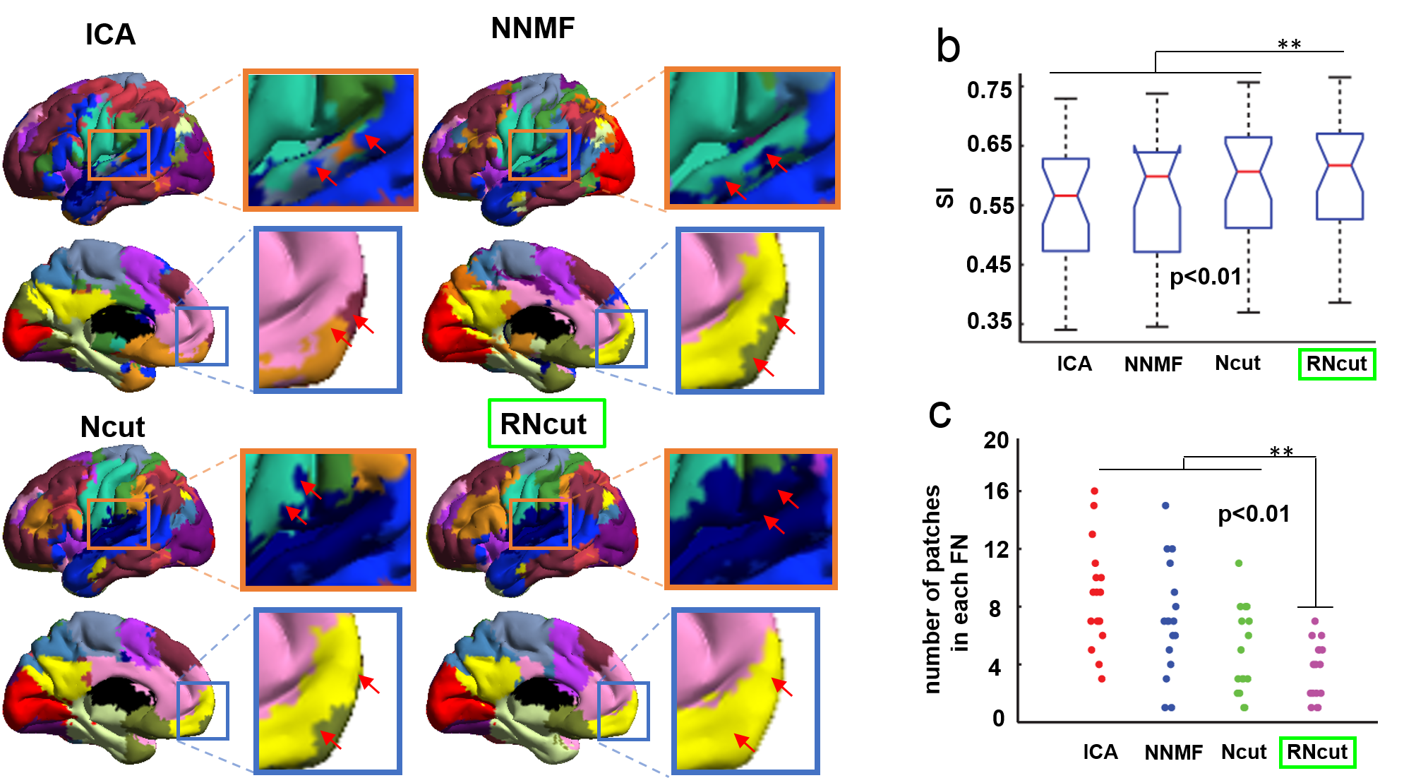
Simulated dataset: We generated the simulated functional dataset using the SimTB toolbox [3] which allowed for flexible generation of fMRI signal time-series (http://mialab.mrn.org/software/simtb/). The dataset consisted of the simulated data of 20 subjects, and each subject had 150 2D images with dimensions of 100x100 voxels, generated by linear combinations of 15 distinct FNs using the following parameter: TR=2000ms with translation and rotation. In addition, Rician noise was added to the simulated images with different contrast-to-noise ratios (CNR). Human subject dataset: Rs-fMRI dataset [4] from 26 subjects with no brain lesions or mental disorders was used for the parcellation evaluation (www.nitrc.org/projects/fcon_1000/). The human subject rs-fMRI images were acquired with a 3T scanner and had the following parameters: resolution = 3.13 × 3.13 × 3.6 mm3, TR = 2s, 225 volumes. Before preprocessing, we removed the first 10 volumes for signal to reach a steady state and kept 215 functional volumes for each subject. Subsequently, we did the slice timing, EPI normalization, spatial smoothing, detrending, temporal filtering, and regression of several nuisance variables. RNcut Method: It was implemented based on the Ncut [7] and added two more constraints including smoothing term (R1) and small patches removal term (R2) as shown in Fig. 1. The R1 term was a Markov random field (MRF) model which made the neighbor voxels share similar cluster labels. The R2 term was to remove the small patches in each functional network. The flowchart of the RNcut was shown in Fig. 2, the weight matrix of each subject was calculated by Pearson correlation from each pair of voxels in the brain within the gray matter mask. The mean weight matrix was obtained by averaging each individual weight matrix. Then, we performed the RNcut on the mean weight matrix to yield the cerebral parcellation. Quantitative evaluation and comparison: The performance of different parcellation methods was evaluated on the simulated data and human subject data. We used the following evaluation measures: (1) Dice and L1 error [8], (2) Clustering validity measures, e.g. Silhouette Index (SI) [2] and the number of patches in each network.Results
Test results with simulated data: Fig 3 shows the parcellation results using different methods on the simulated rs-fMRI data with various levels of noise added. The Fig 3a shows parcellation results using ICA [5], NNMF [6], Ncut [7] and RNcut methods on the simulated rs-fMRI data with low noise level. Fig 3b shows the parcellation results using different methods on the rs-fMRI data with high noise level. Overall, Ncut and RNcut outperformed ICA and NNMF. For rs-fMRI with high noise level, RNcut was more robust to noise while parcellation result from Ncut was significantly affected by noise (Fig 3b). Dice and L1 error measurements in Fig 3c also indicated that RNcut performed best among all methods. Fig 3d shows that smallest number of noise pixels were generated with RNcut. Test results with human subject data: Fig 4a shows the human subject functional networks generated by ICA, NNMF, Ncut and RNcut. Much fewer noisy patches (marked by red arrows) in parcellated functional networks were created by the RNcut method. Fig 4b shows that highest SI value with statistical significance was achieved by the RNcut method. Fig. 4c demonstrates that smallest number of isolated patches were obtained by RNcut.Discussion and Conclusions
We have developed a novel data-driven RNcut method that is more robust to noise and could generate functional networks with higher functional homogeneity and spatial contiguity than widely used ICA, NNMF and Ncut, as demonstrated by the results obtained from both the simulated data (Fig 3) and human subject data (Fig 4). The experimental results demonstrated that RNcut could generate functional networks with higher SI value and less noisy patches. RNcut is computationally efficient and could be used in a wide range of rs-fMRI studies involving functional network parcellation.Acknowledgements
This study is funded by NIH MH092535, MH092535-S1, HD086984 and EB022573.References
[1] Wang, D., et al., 2015. Parcellating cortical functional networks in individuals. Nature neuroscience, 18(12), 1853-1860.
[2] Craddock RC, et al., 2012. A whole brain fMRI atlas generated via spatially constrained spectral clustering. Human Brain Mapping, 33(8), 1914-1928.
[3] Erhardt EB, et al., 2012. SimTB, a simulation toolbox for fMRI data under a model of spatiotemporal separability. NeuroImage 59, 4160-4167.
[4] Biswal BB, et al., 2010. Toward discovery science of human brain function. Proceedings of the National Academy of Sciences,107(10), 4734-4739.
[5] Calhoun VD, et al.,2001. A method for making group inferences from functional MRI data using independent component analysis. Human brain mapping,14(3),140-151.
[6] Anderson A, et al., 2014. Non-negative matrix factorization of multimodal MRI, fMRI and phenotypic data reveals differential changes in default mode subnetworks in ADHD. NeuroImage, 102(1), 207-219.
[7] Shi J, et al., 2000. Normalized cuts and image segmentation. IEEE Transactions on pattern analysis and machine intelligence, 22(8), 888-905. [8] Dice, LR, 1945. Measures of the amount of ecologic association between species. Ecology, 26, 297-302.
[8] Dice, LR, 1945. Measures of the amount of ecologic association between species. Ecology, 26, 297-302.
Figures