5547
Resting Functional Connectivity Across a Language Network Not Related to Task Based Language Laterality Index1Electrical Engineering and Computer Science, Vanderbilt University, Nashville, TN, United States, 2Radiology and Radiological Sciences, Vanderbilt University Medical Center, Nashville, TN, United States, 3Biomedical Engineering, Vanderbilt University, Nashville, TN, United States
Synopsis
Quantification of language dominance via a laterality index is used to assess the risk of language impairment prior to temporal lobe resection, most commonly in temporal lobe epilepsy. While task based functional MRI has become a useful non-invasive method for this measure, its reliability and accuracy can be reduced by poor task performance. In this work, resting fMRI connectivity in a language based network was investigated as an easier and more robust alternative to task based methods. However, results showed poor associations between the resting connectivity and the task based laterality.
Purpose
For patients with temporal lobe epilepsy (TLE), presurgical quantification of language lateralization is important to avoid language impairment after mesial temporal resection. Current guidelines recommend that task based functional MRI (fMRI) be considered as an option for language lateralization in patients with TLE as an alternative to the highly invasive intracarotid amobarbital procedure1. However, task based fMRI studies can be limited by the patient’s ability to efficiently complete the task without error. Resting fMRI does not require the performance of a task and may provide a potential alternative2. This study aims to determine whether resting fMRI using standard regions of interest can yield language lateralization indices highly concordant with tasked based fMRI.Methods
We recruited 40 (36 right handed, 21 males, average age: 40.4 yrs) healthy control subjects with no history of head trauma or neurological or neuropsychological disease, and 23 TLE patients (20 right handed, 12 males, 16 right TLE, 7 left TLE, average age: 23 yrs) who underwent resting and task based fMRI. The MRI images were acquired using a 3T MRI scanner. The functional MRI protocol during the language tasks was as follows: matrix = 80 x 80 voxels, field of view = 240 mm, 34 axial slices, temporal resolution = 2 sec, slice thickness = 3.5 mm/ 0.5 mm gap, 100 volumes, 200 seconds. The same fMRI protocol was used to acquire images at rest with eyes closed, with 300 volumes over 600 seconds. During the task based fMRI the subject alternated between 20 seconds of rest and 20 seconds of silently generating words in a given category. Informed consent was obtained prior to scanning each subject per Vanderbilt University Institutional Review Board guidelines.
Seven regions on interest (ROIs) were identified on each subject: left hippocampus (LHIPP), right hippocampus (RHIPP), left Broca’s Area (LBRO), right Broca’s Area (RBRO), left Wernicke’s Area (LWER), right Wernicke’s Area (RWER), and the supplementary motor area (SMA).
To calculate the task based laterality index (LI), we used the LI Toolbox3. The toolbox uses the number of voxels activated above a certain threshold in the left and right hemisphere ROIs to compute the task based LI. The threshold of activation is determined by a bootstrapping method.
Since there is no current standard for calculating a LI using resting fMRI data, we adopted two different methods to utilize resting data to estimate laterality. Using the resting fMRI images, the average time series for each ROI was computed. The correlation coefficient between each pair of ROIs (n = 21) were computed and converted into z scores. In the first method a resting LI was computed. The correlation coefficients were used in the following equation: LI = (L-R)/(L+R), where L is the correlation coefficient in the left hemisphere such as HIPPL to BROL, and where R is the similar correlation coefficient in the right hemisphere. The correlation between resting and task LIs was computed for different ROIs for all patients and controls together.
The second method to use resting fMRI data compared each pair-wise ROI correlation coefficient (z) with the task based LI to identify any associations between the two. The relationships within the right-handed controls, the left-handed controls, right TLE, and left TLE groups were examined separately.
Results and Discussion
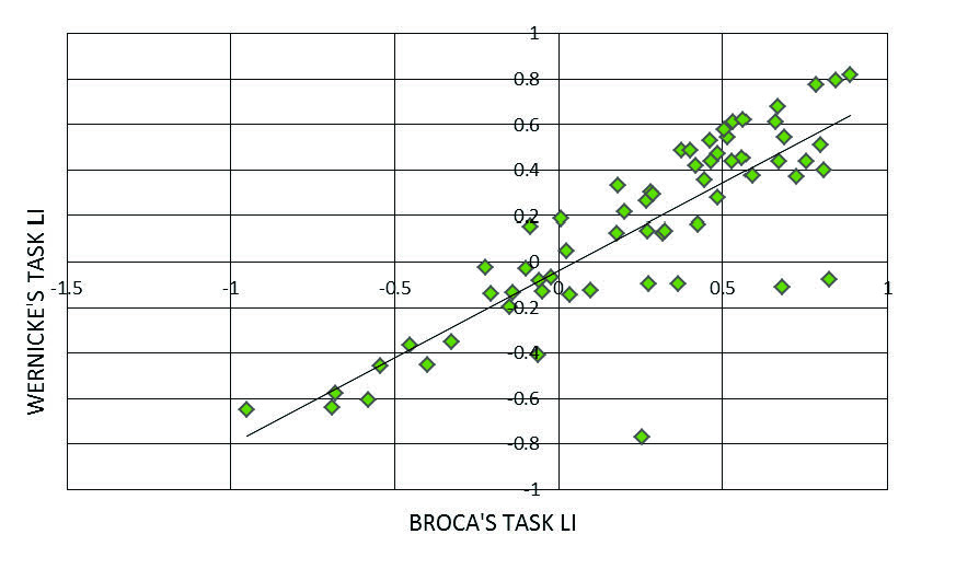
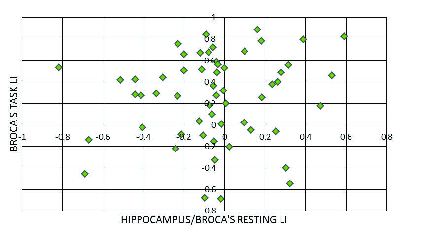
The task based fMRI data computed by the LI Toolbox showed a strong correlation between the LI computed using the Broca’s ROI and Wernicke’s ROI (Figure 1). Future results describe Broca’s ROI. The findings from the first resting method computing the resting LI showed that between the 7 different ROIs, the correlation between the Hippocampus and Broca’s areas presented the greatest overlap with the task LIs. However, overall the resting LI values were generally low suggesting little difference between the resting connectivity in the left and right hemispheres. Therefore, there was no relationship between the resting and task LIs (Figure 2). Other regions showed similar results.
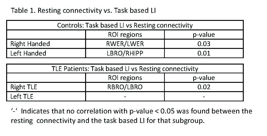
Using the second resting method, the results were similar to the first in that the resting connectivities were not strongly correlated with task based LI. However, subgroup comparisons based on handedness and TLE side showed some weak cross-hemisphere associations between resting connectivity and task based LI (Table 1).
Conclusions
These findings suggest that resting fMRI connectivity across the language network identified here is not an alternative to the task-based laterality indices. Further studies may focus on examining the weak correlations between cross-hemisphere ROI pairs. This study has not invalidated the use of resting fMRI to compute LI, but more development is required.Acknowledgements
This work was supported by NIH R01 NS75270 (Morgan)References
1. Jerzy P. Szaflarski, et al., “Practice guideline summary: Use of fMRI in the presurgical evaluation of patients with epilepsy: Report of the Guideline Development, Dissemination, and Implementation Subcommittee of the American Academy of Neurology,”Neurology, vol. 88, pp. 395-402, Jan. 2017.
2. Rogers B. P., Morgan V. L., Newton A. T., Gore J. C, “Assessing functional connectivity in the human brain by fMRI,” Magn. Reson. Imaging, vol. 25, pp. 1347–1357, May 2007.
3. https://www.medizin.uni-tuebingen.de/kinder/en/research/neuroimaging/software/
Figures